Figures & data
Figure 1. Experimental protocols of the study. (A) Study with dinitrochlorobenzene (DNCB) and acetic acid (AA)-induced ulcerative colitis rats; (B) study with bacterial translocation of colitis rats.
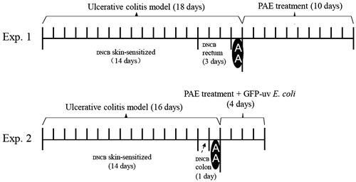
Table 1. Evaluation of disease activity index (DIA).
Table 2. Ingredients of the three fractions of PAE.
Figure 2. Macroscopic appearances of normal rat rectum (A) and dinitrochlorobenzene and acetic acid-induced colitis rat rectum (B).
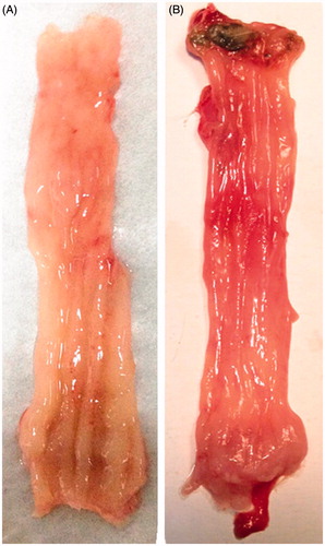
Figure 3. Enemas administration of P. americana extract (PAE) ameliorated dinitrochlorobenzene and acetic acid-induced colitis. Changes in disease activity index score were determined daily throughout the 10-d experiment period.
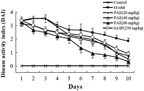
Figure 4. Representative H&E staining photomicrographs on rats rectum sections. (A) The vehicle group shows intact crypts structure and epithelial cell layer, normal goblet cells morphology. (B) The model group shows lamina propria damage, epithelial cells loss and intense inflammatory cells infiltration. (C) The sulphasalazine (SASP) (250 mg/kg) group shows regenerated mucosa and crypts and decreased inflammatory reaction. (D) The P. americana extract (PAE) (80 mg/kg) group shows no remarkable inflammatory features with normal crypts structure. (E) The PAE (40 mg/kg) group shows crypts dilation and distortion, fibrosis in lamina propria. (F) The PAE (20 mg/kg) group shows disappearance of crypts, superficial mucosal ulcer and inflammatory cells infiltration in the lamina propria. Magnification was 100×.
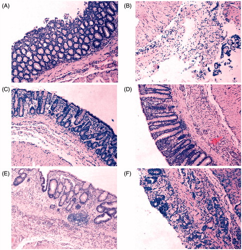
Figure 5. MPO levels of rectum segments from rats treated with P. americana extract (PAE) (20, 40 and 80 mg/kg/day enemas) or sulphasalazine (SASP) (250 mg/kg/d enemas). +++ p < 0.001 versus the vehicle group; ***p < 0.001 versus the dinitrochlorobenzene and acetic acid-induced colitis group.
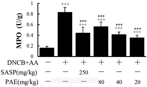
Figure 6. A cultured sample of fluorescent image of GFP-uv E. coli isolated from ulcerative colitis rat tissue under an ultraviolet lamp at 540 nm.
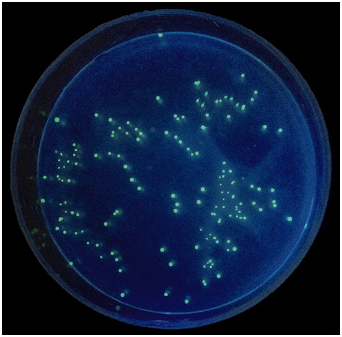
Table 3. CFU number of viable GFP-uv E. coli and incidence of bacterial translocation of rats tissues. (n = 6/group).
Figure 7. Effects of P. americana extract (PAE) and its three fractions on proliferation of NIH 3T3 fibroblasts. *p < 0.05, **p < 0.01, ***p < 0.001 versus the control group. #p < 0.05 versus the PAE group.
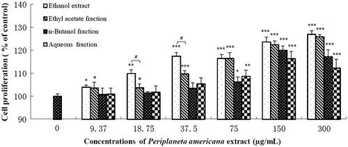
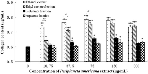
Figure 8. Effects of P. americana extract (PAE) and its three fractions on collagen production of NIH 3T3 fibroblasts. *p < 0.05, **p < 0.01, ***p < 0.001 versus the control group. #p < 0.05, ##p < 0.01 versus the PAE group.