Figures & data
Figure 1. Bronchial mucosa blood flow (BMBF) with laser-Doppler velocimetry. Left panel: Left BMBF in study and control group. Right panel: Left/right BMBF ratio in study and control group. Filled circle: study group. Open circle: control group.
Table I. Overview of results.
Figure 2. Bronchial mucosa haemoglobin saturation with DRS. Left panel: Bronchial mucosa saturation of left bronchus in study and control group. Right panel: Left/right ratio of bronchial mucosa saturation in study and control group. Filled circle: study group. Open circle: control group.
Figure 3. Bronchial mucosa haemoglobin concentration with DRS. Left panel: Bronchial mucosa con-centration of left bronchus in study and control group. Right panel: Left/right ratio of bronchial mucosa concentration in study and control group. Filled circle: study group. Open circle: control group.
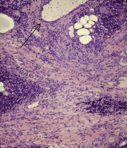
Figure 4. Section of left lung with major bronchi (H&E, × 100), showing severe broadening of lung septae with inflammation, oedema and slight fibrosis. Specimen was taken peripheral to both anastomosis and measuring site. Arrow: Small bronchus.