Figures & data
Table I. General characteristics of participants.
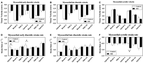
Figure 1. Tissue Doppler Imaging of left anterior descending artery (LAD) supplied left ventricular segments (17 segments model) in patients with and without mid-LAD myocardial bridge. The bar above each column indicates standard error of mean. A to C shows tissue velocity of 7 LAD-specific segments, and D to F show the corresponding strain rate values. MB, myocardial bridge; Apical-L, apical lateral; Basal-A, basal anterior; Mid-A, mid anterior; Apical-A, apical anterior; Basal-AS, basal anteroseptal; Mid-AS, mid anteroseptal; Apical-S, apical septal. *P < 0.05.

Figure 2. Relationships between myocardial bridge angiographic features [stenosis (A) and length (B)], their interactions (C) and left ventricular systolic synchrony. The dash line started from Y axis indicates the cutoff value of 1.91% derived from control group to define the presence of LV systolic dyssynchrony. Solid line in each scatter plot indicating the median. Tmsv-16SD/RR, the time-to-minimum systolic volume corrected by RR interval. SDI, systolic dyssynchrony index. *P < 0.05, **P < 0.01, ***P < 0.001.
![Figure 2. Relationships between myocardial bridge angiographic features [stenosis (A) and length (B)], their interactions (C) and left ventricular systolic synchrony. The dash line started from Y axis indicates the cutoff value of 1.91% derived from control group to define the presence of LV systolic dyssynchrony. Solid line in each scatter plot indicating the median. Tmsv-16SD/RR, the time-to-minimum systolic volume corrected by RR interval. SDI, systolic dyssynchrony index. *P < 0.05, **P < 0.01, ***P < 0.001.](/cms/asset/7728e3c8-877e-4a7d-b051-119630f69874/icdv_a_736635_f0002_b.gif)
Figure 3. Interaction of myocardial bridge percent stenosis and length on trans-mitral E wave peak velocity (A) and E/A ratio (B). Solid line in each scatter plot indicating the median. MB, myocardial bridge. *P < 0.05.

Figure 4. Receiver operator characteristic curve analysis of trans-mitral E wave peak velocity (A), E/A ratio (B) and myocardial bridge stenosis (C) for the diagnostic value of left ventricular dyssynchrony detected by real-time three dimensional echocardiography. AUC, area under curve; CI, confidence interval; MB, myocardial bridge.

Table II. Univariate logistic regression analyses.
Table III. Multivariate logistic regression analyses.