Figures & data
Table I. Comparison of fibrosis parameters between days 9 and 14 within each experimental group.
Figure 1. Representation of fibrosis distribution and severity parameter scores at days 9 and 14 post-intratracheal bleomycin instillation. (A–D) Fibrosis distribution (A) and severity (B–D) at day 9. (E–H) Fibrosis distribution (E) and severity (F–H) at day 14. (A, E) Fibrosis distribution; (B, F) fibroblast proliferation; (C, G) collagen deposition; (D, H) alveolar obliteration. The number of mice in each group is indicated (n). Brackets represent significant differences between groups; *P < 0.05; **P < 0.01.
Figure 2. Digital image morphometric analysis of lung collagen deposition. (A) Microphotographs taken before (left panel) and after (right panel) the imaging analysis procedure used to quantitate the collagen staining (black areas) from the image. The identification number of each mouse is indicated on each microphotograph. (B) Median values with IQR of quantitative collagen deposition are represented as box-plots for Bleo (dark gray), Bleo + non-CM (light gray) and Bleo + AMTC-CM (white) groups. The number of mice in each group is indicated (n). Brackets represent significant differences between groups; *P < 0.05.
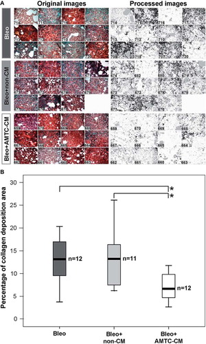
Figure 3. Representation of overall fibrosis score at days 9 and 14 post-intratracheal bleomycin instillation. Box-plots reporting the median and IQR of values obtained from the Bleo (dark gray), Bleo + non-CM (light gray) and Bleo + AMTC-CM (white) groups are represented for each time-point. The number of mice in each group is indicated (n). Brackets represent significant differences between groups and time-points; *P < 0.05; **P < 0.01.