657
Views
2
CrossRef citations to date
0
Altmetric
Research Article

Biotransformation studies of prednisone using human intestinal bacteria Part II: Anaerobic incubation and docking studies

, , &
Pages 1211-1219 | Received 28 Nov 2008, Accepted 20 Jan 2009, Published online: 16 Nov 2009

Figures & data

Figure 1. Separation of prednisone metabolites (3–9) after anaerobic incubation with human intestinal bacteria (HIB).
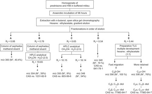
Figure 1.  Separation of prednisone metabolites (3–9) after anaerobic incubation with human intestinal bacteria (HIB).

Table 1. 13C-NMR assignments of prednisone 1 and its anaerobic metabolites 3–9.

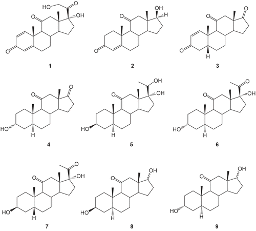
Figure 2. Chemical structures of prednisone 1, testosterone 2, and HIB metabolites of 1 (3–9).

Figure 2.  Chemical structures of prednisone 1, testosterone 2, and HIB metabolites of 1 (3–9).

Figure 3. The steroid-binding site of PGE2 9-ketoreductase. Structures of PGE2 9-KR (taken from PGE2 9-KR, NAD,·testosterone complex, light gray). Residues that contact the steroid ligands such as Phe 54, Tyr 55, His 117, Phe 118, Ile 129, Val 306, and Phe 310 are shown in stick representation.
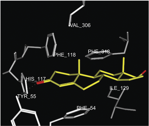
Figure 3.  The steroid-binding site of PGE2 9-ketoreductase. Structures of PGE2 9-KR (taken from PGE2 9-KR, NAD,·testosterone complex, light gray). Residues that contact the steroid ligands such as Phe 54, Tyr 55, His 117, Phe 118, Ile 129, Val 306, and Phe 310 are shown in stick representation.

Figure 4. Docking of metabolite 4 (yellow stick) in the active site of PGE2 9-KR. Hydrogen atoms of the amino acid residues have been removed to improve clarity.
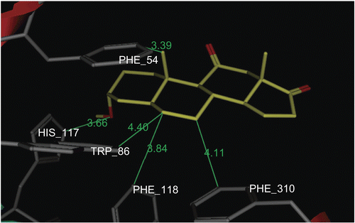
Figure 4.  Docking of metabolite 4 (yellow stick) in the active site of PGE2 9-KR. Hydrogen atoms of the amino acid residues have been removed to improve clarity.
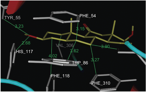
Figure 5. Docking of metabolite (yellow stick) in the active site of PGE2 9-KR. Hydrogen atoms of the amino acid residues have been removed to improve clarity.

Figure 5.  Docking of metabolite 8β (yellow stick) in the active site of PGE2 9-KR. Hydrogen atoms of the amino acid residues have been removed to improve clarity.

Figure 6. Docking of metabolite (yellow stick) in the active site of PGE2 9-KR. Hydrogen atoms of the amino acid residues have been removed to improve clarity
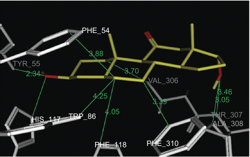
Figure 6.  Docking of metabolite 8α (yellow stick) in the active site of PGE2 9-KR. Hydrogen atoms of the amino acid residues have been removed to improve clarity

Table 2. Results of PASS predicted potential and molecular modeling of prednisone metabolites (3–9).
Reprints and Corporate Permissions

Please note: Selecting permissions does not provide access to the full text of the article, please see our help page How do I view content?

To request a reprint or corporate permissions for this article, please click on the relevant link below:

Academic Permissions

Please note: Selecting permissions does not provide access to the full text of the article, please see our help page How do I view content?

Obtain permissions instantly via Rightslink by clicking on the button below:

If you are unable to obtain permissions via Rightslink, please complete and submit this Permissions form. For more information, please visit our Permissions help page.