Figures & data
Figure 1. Chemical structure of acetazolamide (AZM) exists in each monomer.

Figure 2. A snapshot picture taken from the simulation box: Protein (CA IX-dimer in yellow, inhibitor (inside the protein), water (as quick surface) and K+, Cl− ions (cyan and brown colored spheres).
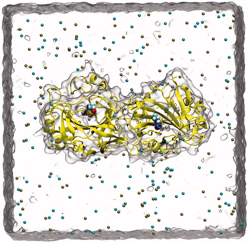
Figure 3. Cognate docking poses alignment with the co-crystallized ligand conformation using different methods.
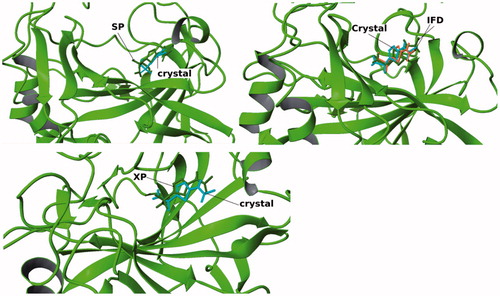
Figure 4. Superimposition of selected ligands from docking simulations in the active site.
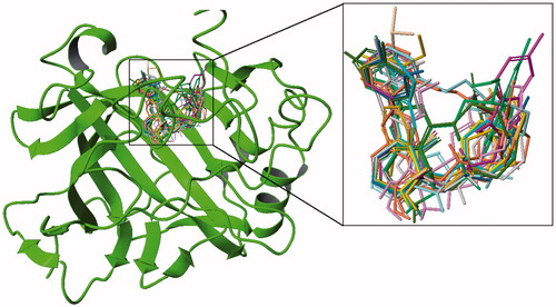
Table 1. 2D chemical structures and their corresponding docking scores for selected top-docking scored compounds.
Figure 5. 3D and 2D ligand interaction diagrams for the ligands, ZINC20464003 (upper panel) and ZINC72421916 (lower panel).
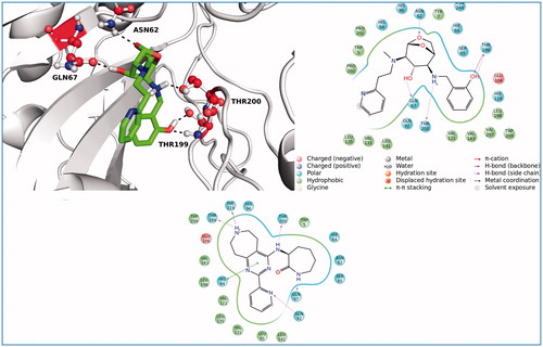
Table 2. Selected compounds with their ADME properties.
Figure 6. 3D docking poses of selected compounds at the active site of CA-IX.
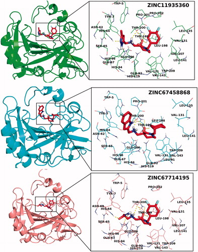
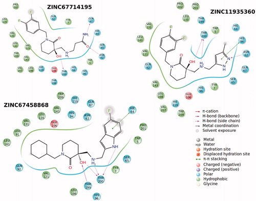
Figure 7. 2D ligand interaction diagrams of selected compounds into the active site of CA-IX.