Figures & data
Figure 1. Chemical structures of the isolated compounds (1–6) and ursolic acid (7).
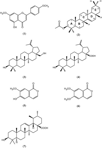
Table 1. PTP1B activity of A. roxburghiana compounds and ursolic acid*.
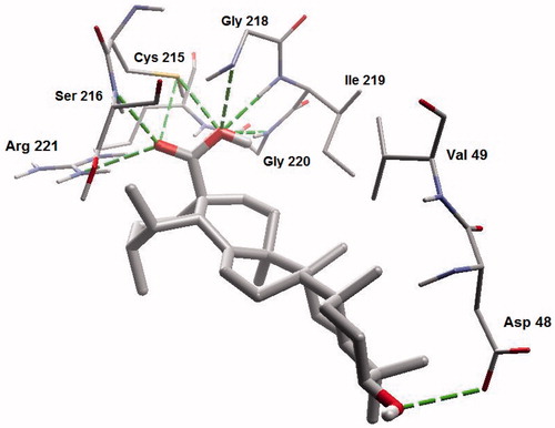
Figure 2. Molecular binding mode of betulin inside catalytic site of PTP13. Hydrogen atoms (except polar ones) were omitted for clarity.
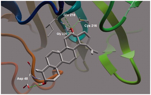
Figure 3. A closer view of betulin bound to the active site of PTP13.

Figure 4. Electrostatic surface of PTP1B. Blue color indicates positive electrostatic surface, red color region indicates negative electrostatic surface and white region depicts non-polar (hydrophobic) surface of the enzyme.
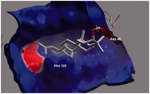
Figure 5. Similar binding modes of betulin and betulic acid. Both poses of the compounds seems to be superimposed on each other. It clearly depicts similar bonding interactions between the enzyme and inhibitors.
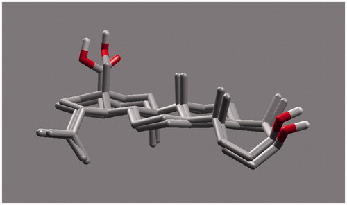
Figure 6. Molecular binding mode of ursolic acid inside active site of PTP13.
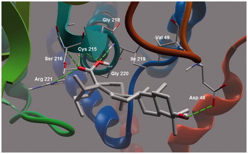
Figure 7. A closer view of ursolic acid bound to the active site of PTP1B.