Figures & data
Table 1. Structures of 2a–j and 3d–i (these compounds were available from previous studies).
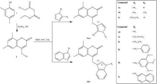
Scheme 1. Synthesis of 6,8-dimethyl coumarin bearing imidazolium and benzimidazolium salts (1a–i).
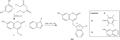
Scheme 2. Synthesis of 7-hydroxy coumarin bearing benzimidazolium salts (2g, h).
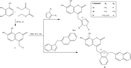
Scheme 3. Synthesis of 7,8-dihydroxy coumarin bearing imidazolium and benzimidazolium salts (3a–c, j).
Scheme 4. Synthesis of benzoxazinone bearing imidazolium and benzimidazolium salts (4a–i).
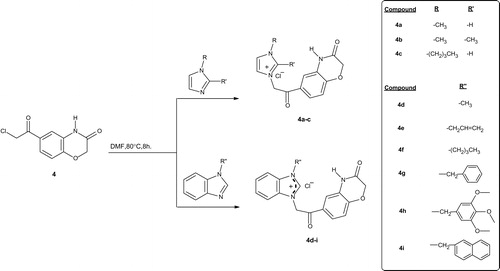
Table 2. IC50 values of synthesized and previously reported compounds against hCA I and II inhibition.
Table 3. ScMet and neurotoxicity screening data in mice dosed ip with the compounds.
Table 4. MES screening data of 1i, 2f, 3g and 3j in mice dosed ip.
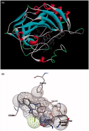
Figure 1. Docking of compound 3j within the hCA II active site. (A) Discovery Studio 4.0 Client images and (B) ADT images.
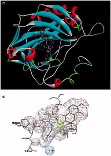
Figure 2. Docking of compound 1i within the hCA II active site. (A) Discovery Studio 4.0 Client images and (B) ADT images.