Figures & data
Figure 1. (a) Novel minimally invasive radiofrequency device. (b) Procedure: (Citation1) Insertion of the needle into the scar at variable depths from the superficial to mid-dermis (Citation2) ‘in situ’ 360 degree rotation (tunneling) performed to create a virtual canal contextually supplying the radiofrequency (Citation3) Repetition of the procedure in retrograde using a multi-prick technique along the scar.
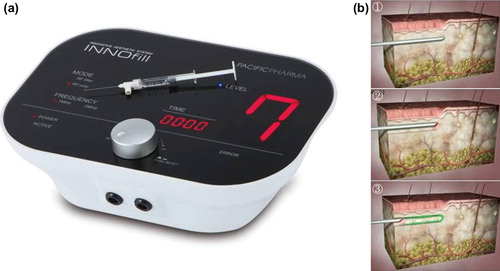
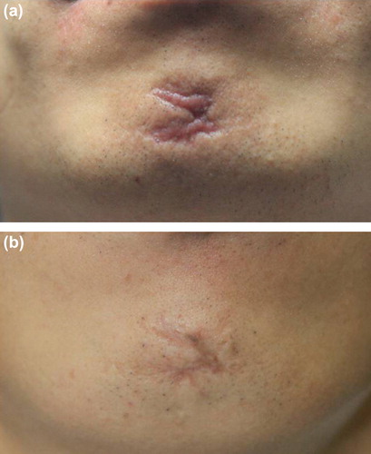
Figure 2. (a) Depressed, curved facial scar on the mid-chin and (b) marked improvement 2 months after intradermal RF treatment.