Figures & data
Figure 1. a) Transabdominal ultrasonographic image at 27 weeks of gestation showing the monochorionic-diamniotic placenta with discordant echogenicity; the hyperechogenic, thicker portion belonged to the donor twin (left), whereas the placental side of the recipient twin had a normal echotexture (right). b) Color Doppler examination showing the presence of blood flow in the depth of the placenta in the recipient (R) side. In contrast, reduced vascular Doppler signals were observed in the donor (D) side.
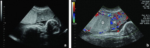
Figure 2. Maternal surface of the monochorionic-diamniotic placenta showing stark contrast between the donor (D) and the recipient (R) sides. The donor side is more pale and shaggy, while the recipient side is dark red, congested appearance.
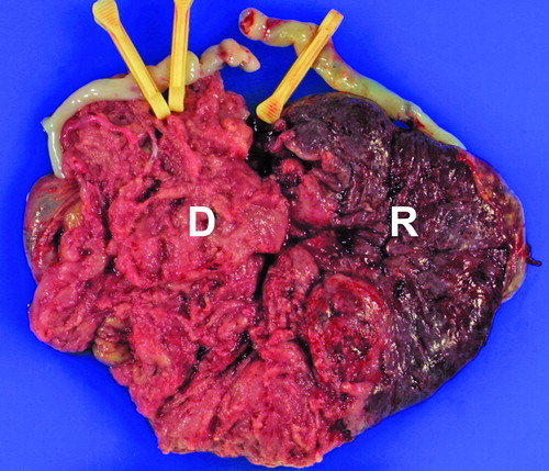
Figure 3. Histologic examination of the placenta with hematoxylin and eosin (H&E): (A) The donor side shows alternating areas of edematous or fibrotic immature intermediate villi; (B) The maturation pattern of chorionic villi of the recipient side is uniform and is compatible with that of a third trimester placenta.
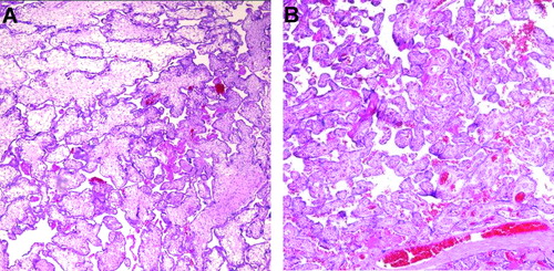
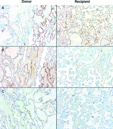
Figure 4. Protein expression of placental growth factor (PlGF), VEGF receptor-1 (VEGFR-1), and Endoglin in the monochorionic-diamniotic placenta. The donor side is characterized by variable expression of these molecules: (A) PlGF was expressed in both the donor and the recipient tissues, but the immunoreactivity was variable in the donor side, edematous villi being weakly reactive. In contrast, moderate to high distinct immunoreactivity for (B) VEGFR-1 and (C) Endoglin was found in the villous trophoblast of the donor side, while the recipient tissue showed decreased or lack of immunoreactivity.