Figures & data
Figure 1. Eosinophils in the amniotic fluid. Amniotic fluid obtained from a patient with spontaneous preterm labor and intact membranes. The eosinophils (arrows) were the predominant cells in the amniotic fluid (arrowhead: epithelial cell).
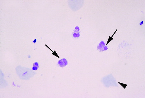
Table I. Clinical characteristics of the study population.
Figure 2. Amniocentesis-to-delivery interval according to the presence or absence of amniotic fluid white blood cell count differential of more than 20% of eosinophils. Although the difference was not significant, the median amniocentesis-to-delivery interval was 7 days shorter in cases than controls [cases: 34 days (95% CI 0–70 days) vs. controls: for cases and 41 days (95% CI 34–48 days); p = 0.084]. Solid line: cases; dashed line: controls; open circles: censored patients.
![Figure 2. Amniocentesis-to-delivery interval according to the presence or absence of amniotic fluid white blood cell count differential of more than 20% of eosinophils. Although the difference was not significant, the median amniocentesis-to-delivery interval was 7 days shorter in cases than controls [cases: 34 days (95% CI 0–70 days) vs. controls: for cases and 41 days (95% CI 34–48 days); p = 0.084]. Solid line: cases; dashed line: controls; open circles: censored patients.](/cms/asset/15575a5d-6e96-451c-9b96-f46ee7016bb1/ijmf_a_417019_f0002_b.jpg)