Figures & data
Table 1. Subject characteristics
Figure 1. Pre- and post-bronchodilator static lung volumes are shown for each GOLD stage group compared with an age-matched healthy control group. Residual volume (RV) and functional residual capacity (FRC) increased progressively as GOLD stage worsened. Total lung capacity increased in GOLD stage I in conjunction with a preserved inspiratory capacity (IC), vital capacity (VC) and expiratory reserve volume (ERV). IC and VC then decreased progressively in GOLD stages II and III/IV.
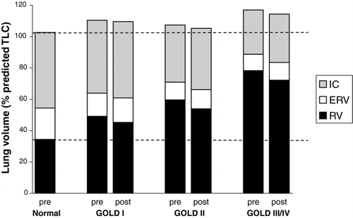
Figure 2. Relationships between forced vital capacity (FVC), residual volume (RV), diffusing capacity of the lung (DLCO) and specific airway resistance (sRaw) are shown against FEV1 (all measurements expressed as% of predicted normal values). FVC changed linearly with FEV1, RV and sRaw both increased exponentially as FEV1 decreased, and DLCO decreased as FEV1 decreased.
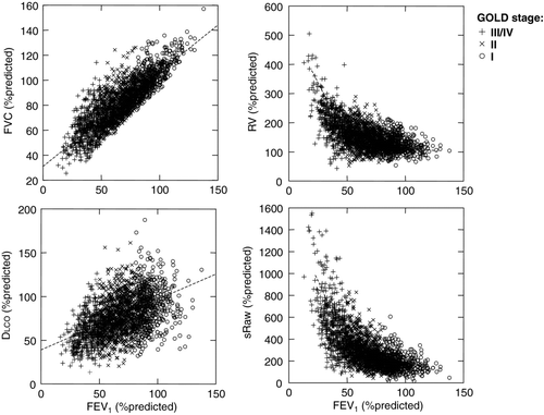
Table 2. Bronchodilator reversibility: pre- to post-bronchodilator changes
Figure 3. Bronchodilator-induced changes in lung volumes are shown as (A) measured changes expressed as % of predicted normal and (B) frequencies of a positive response defined as at least a 10 % of predicted improvement in that measurement. As GOLD stage worsened, the FEV1 response became less important while other lung volume responses increased progressively.
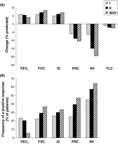
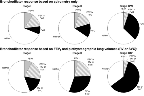
Figure 4. The frequency of a significant bronchodilator response (change of at least 10% predicted) is shown when only spirometric measurements are taken into account (FEV1 alone, FVC alone or combination of FEV1 and FVC) (top). By considering plethysmographic lung volume (RV and SVC) improvements in addition to spirometric FEV1, greater rates of reversibility were uncovered (bottom). When volumes were taken into account, the frequency of a positive bronchodilator response increased with worsening GOLD stage.