Figures & data
Table 1. Characteristics of patients
Figure 1. Box plots of the mean physical activity for the entire measurement period assessed at ankle and wrist (I) and hip (II) in patients with (group A) and without (group B) walkers.
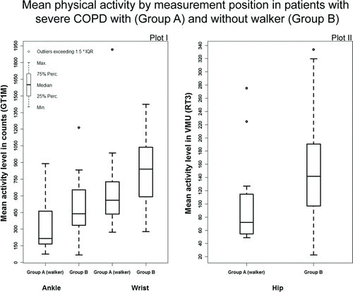
Table 2. Measurements of activity obtained by the GT1M (wrist, ankle) and the RT3 (hip) in patients with and without walker
Table 3. Correlations between activity data from different monitor positions in patients with and without walker
Figure 2. Relationship between the mean PA values assessed at the ankle and the 6MWD assessed at the beginning and end of the PR program in both patient groups.
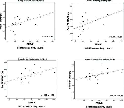
Table 4. Standard multivariate regression model to assess the impact of group and gender and all simultaneously measured PA positions, on the disease severity as described by the BODE score