Figures & data
Figure 1. Disposition process for patient selection.
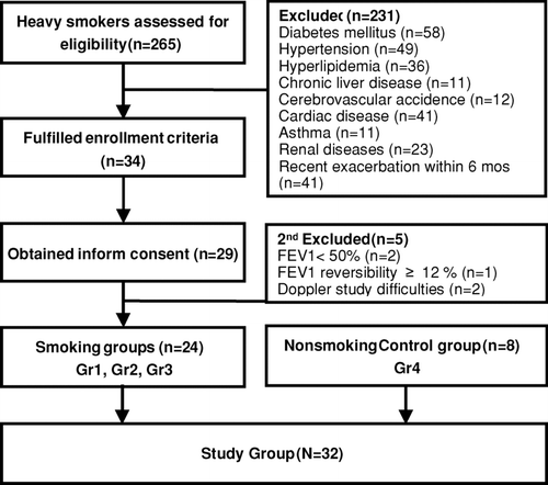
Table 1. Clinical characteristics, arterial blood gas analysis, and pulmonary function of the study population*.
Figure 2. Renal resistive index before and after protein loading in smoker patients with different severities of COPD and nonsmoker control subjects. The postprandial RRIs were significantly increased compared with the baseline values in the heavy smoking patients. Moderate COPD patients (FEV1 50–80% predicted, Group 3) had persistent RRI elevation for more than 90 minutes, and the RRI elevation lasted for only 45 minutes in the other two smoker groups. Group 1: smokers with normal spirometry, 8 patients; Group 2: mild COPD, 8 patients; Group 3: moderate COPD, 8 patients; Group 4: nonsmoker control group, 8 subjects. *p < 0.05,**p < 0.01 compared with baseline RRI by paired t-tests. § p < 0.05 compared with Group 1 at the same time points by the Student's t-test.
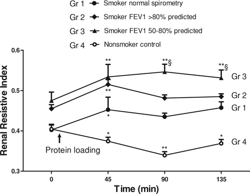
Figure 3. Renal resistive index before and after protein loading in patients with high/low serum TNF-α levels. The RRIs were significantly increased from the baseline values in patients with high TNF-α (> 7 pg/ml) and low TNF-α (≤ 7 pg/ml) levels 45 min postprandial, while the RRI elevation lasted longer in patients with high TNF-α levels (*p < 0.05,**p < 0.01 compared with baseline RRI by paired t-tests). The RRI was also significantly more elevated in patients with high TNF-α levels compared with those with low TNF-α levels at 135 min (§ p < 0.05 by the Student's t-test).
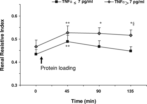
Figure 4. Renal resistive index 135 min after protein loading against airway obstruction and TNF-α serum levels. (A) The RRI was negatively correlated with the percentage of predicted values of FEV1 (Correlation Coefficient = –0.563, P = 0.010). (B) The RRI was positively correlated with serum TNF-α levels (Correlation Coefficient = 0.473, P = 0.035). The solid lines indicate the regression line and the dashed lines indicate the 95% confidence intervals.
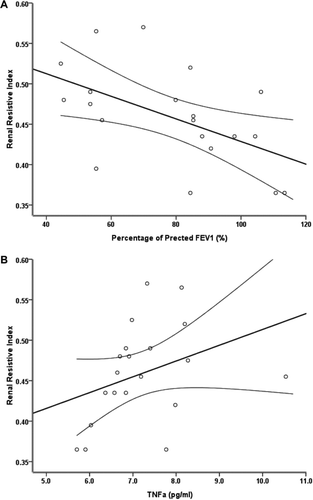
Table 2. Multiple linear regression of the final renal resistive indices.