Figures & data
Table 1. Anthropometric clinical, functional and imaging data (mean ± SD) or n (%) of participants by ATS/ERS stages of disease severity
Figure 1. Prevalence of the different DRB1 (top panel) and DQB1 (bottom panel) allelic frequencies, ordered from most to less prevalent in controls (white columns), compared to those determined in patients with COPD (grey columns).
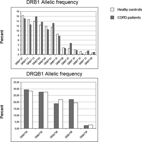
Table 2. Number of DRB1 and DQB1 alleles (n), allelic frequency (AF%) and Odds Ratio (OR) in controls and COPD patients
Table 3. Haplotype frequency (HF%) of HLA DRB1-DQB1 haplotypes in COPD patients
Table 4. Number of alleles (n) and DRB1 allelic frequency (AF%) in COPD patients by severity of airflow limitation (ATS/ERS stages of disease severity)
Table 5. Number of alleles (n) and DRB1 allelic frequency (AF%) in COPD patients by DLCO impairment