Figures & data
Table 1. Demographic and clinical characteristics of the subjects
Table 2. Total and differential cell counts of mini-BAL
Table 3. Microbiology of mini-BAL
Figure 1. Increased expression of TLR4 in mini-BAL but not in the peripheral blood of S and of COPD. Mini-BAL cells and paired blood samples were recovered from C (n = 10), from S (n = 8) and from COPD (n = 18) patients. The expression of TLR4 was assessed by immunocytochemistry using an anti-TLR4 polyclonal antibody; *p < 0.05 vs C; **p < 0.05 vs S; #p < 0.05 vs autologous peripheral blood. Data are expressed as median (25-75 percentiles).
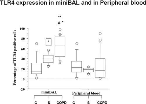
Table 4. Total and differential cell counts of mini-BAL
Figure 2. Absence of differences in the concentrations of IL-8 in mini-BAL and in the peripheral blood of S and of COPD. Mini-BAL supernatants and paired blood samples were recovered from C (n = 10), from S (n = 8) and from COPD (n = 18) patients. IL-8 concentrations were measured by ELISA as described in “materials and methods” and are expressed as pg/ml. Data are expressed as median (25-75 percentiles).
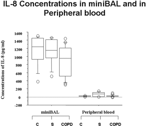
Figure 3. Reduced concentrations of IP-10 in mini-BAL but not in the peripheral blood of COPD. Mini-BAL supernatants and paired blood samples were recovered from C (n = 10), from S (n = 8) and from COPD (n = 18) patients. IP-10 concentrations were measured by ELISA as described in “materials and methods” and are expressed as pg/ml. *p < 0.05 vs C; **p < 0.05 vs S; #p < 0.05 vs autologous peripheral blood. Data are expressed as median (25-75 percentiles).
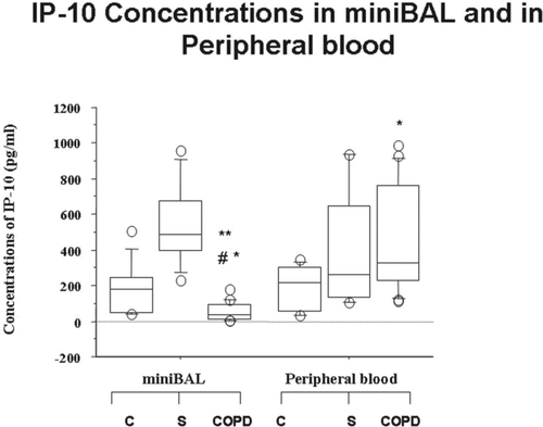
Figure 4. Reduced expression of Foxp3 in mini-BAL and in the peripheral blood of S and of COPD. Mini-BAL cells and paired blood samples were recovered from C (n = 10), from S (n = 8) and from COPD (n = 18) patients. Total proteins were extracted and analysed for Foxp3 expression by western blot analysis. Membranes were then stripped and incubated with goat polyclonal anti–ß-actin. A. Densitometric analysis of Foxp3 expression. Signals corresponding to Foxp3 on the various western blots were semiquantified by densitometric scanning, normalized and expressed after correction with the density of the band obtained for beta-actin (mean±SD). * p < 0.05. B. Representative Western blot analysis for Foxp3 expression from C, S and COPD subjects. Data are expressed as median (25-75 percentiles).
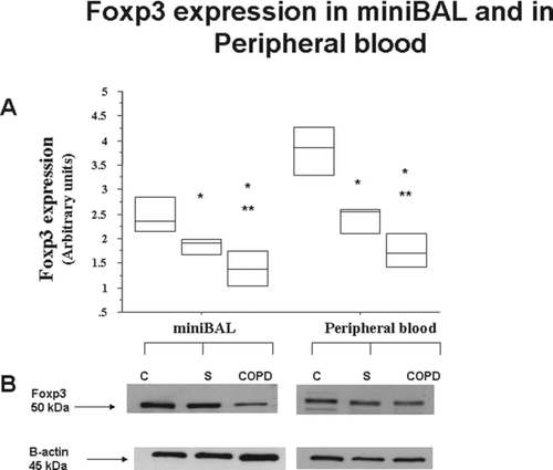
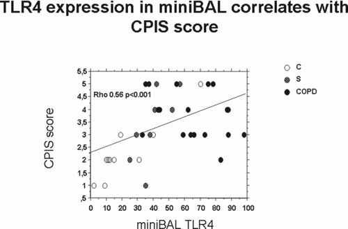
Figure 5. Correlations between the expression of TLR4 in mini-BAL and CPIS score. The expression of TLR4 in mini-BAL of C (n = 10), S (n = 8) and COPD (n = 18) was correlated with CPIS score by Spearman Correlation test.