Figures & data
Figure 1. Measured values, from arterial blood gases, from patients 1 to 6, for wash out (circles) and wash in (stars) of PO2 (1a to 6a) and SO2 (1b to 6b), plus single exponential curves fitted to measured values.
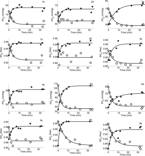
Figure 2. Measured values from arterial blood gases from patients 7 to 12, for wash out (circles) and wash in (stars) of PO2 (7a to 12a) and SO2 (7b to 12b), plus single exponential curves fitted to measured values.
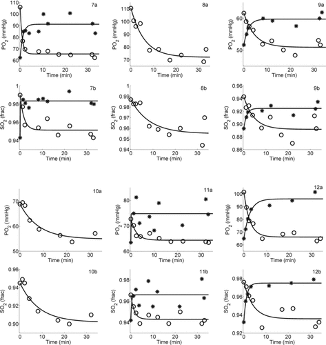
Table 1a. Oxygen administered, acid-base and oxygenation status in the 12 patients at the start and the end of oxygen wash out
Table 1b. Oxygen administered, acid-base and oxygenation status in the 12 patients at the start and the end of oxygen wash in and 30 minutes following the end of wash in.
Table 2. Lung function, time constants (τ) and time to clinical steady state (Tp, Ts) for oxygen wash out and wash in, in each of the 12 patients
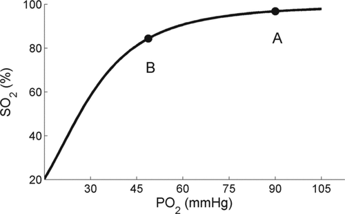
Figure 3. The oxygen dissociation curve (ODC). A and B are used in the text to describe changes in oxygenation from these points.