Figures & data
Table 1. Characteristics of study patients
Table 2. qPCR detection of respiratory viruses
Table 3. Quantitative histology for airway inflammatory cells*
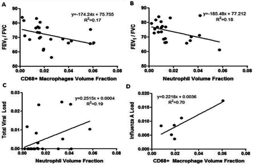
Figure 1. Linear regression analysis of the relationship between inflammatory cells and lung function, and viral load. A) FEV1/FVC vs. Volume fraction CD68 + macrophages (p = 0.044). B) FEV1/FVC vs. Volume fraction neutrophils (p = 0.047). C) Total viral load (normalized to RPP) vs. Volume fraction of neutrophils (p = 0.031). D) Influenza A viral load (normalized to RPP) vs. Volume fraction of CD68+ macrophages (influenza A-positive cases; p = 0.038).