Figures & data
Table 1. Demographic and lung function tests data
Figure 1. The 5-minute averaged heart rate (beat per minute, bpm) in patients and controls (ANOVA p < 0.0001).
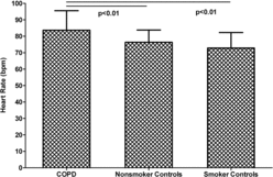
Figure 2. HRV parameters (SDNN, RMSDD, total power, LF modulation and HF modulation) in patients and controls. SDNN: standard deviation of all the NN intervals; RMSSD: root mean square of successive differences between adjacent NN intervals; LF modulation: power in the low frequency band, i.e., 0.04–0.15 Hz; HF modulation: power in the high frequency band, i.e., 0.15–0.4 Hz; Units: SDNN and RMSDD in ms and total power, LF modulation and HF modulation in ms2; values are natural log- transformed; *p < 0.05, ***p < 0.001, + 0.05 > p < 0.10.
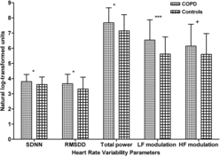
Table 2. Heart rate variability indices in the three groups
Table 3. Heart rate and heart rate variability indices by GOLD severity and by quartiles of BODE index scores
Figure 3. Relationship between heart rate and single-breath diffusion capacity for carbon monoxide (DLCOSB% predicted).
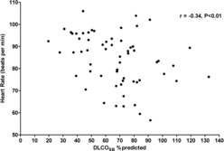
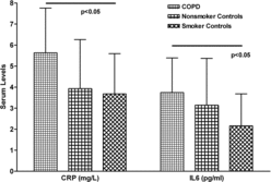
Figure 4. Levels of serum C-reactive protein (CRP) expressed in mg/L, and interleukin 6 (IL-6) expressed in pg/ml, in patient and control groups. (ANOVA p < 0.05 for both).