Figures & data
Table 1. Demographic and clinical data
Table 2. Serum concentration of the L-arginine pathway metabolites and hsCRP in patients with COPD
Figure 1. Serum ADMA levels in COPD patients with and without acute exacerbation compared to healthy subjects. Serum concentration of ADMA (μmol/l) is shown in COPD patients with acute exacerbation (AECOPD), stable COPD and in healthy subjects (HS). Data are presented as mean and 95% confidence interval.
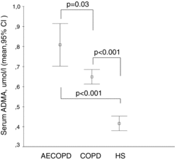
Figure 2. Serum SDMA levels in COPD patients with and without acute exacerbation compared to healthy subjects. Serum concentration of SDMA (μmol/l) is shown in COPD patients with acute exacerbation (AECOPD), stable COPD and in healthy subjects (HS). Data are presented as mean and 95% confidence interval.
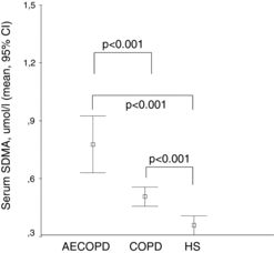
Figure 3. Serum L-arginine levels in COPD patients with and without acute exacerbation compared to healthy subjects. Serum concentration of L-arginine (μmol/l) is shown in COPD patients with acute exacerbation (AECOPD), stable COPD and in healthy subjects (HS). Data are presented as mean and 95% confidence interval.
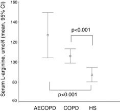
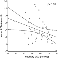
Figure 4. Correlation between partial capillary oxygen pressure and ADMA in patients with COPD. Correlation between partial capillary oxygen pressure (mmHg) and serum concentration of ADMA (μmol/l) is shown in COPD patients. (Pearson correlation, r = -0.34, p < 0.05, scatter plot diagram).