Figures & data
Figure 1. Western blotting and RT-PCR analysis of TLR3 expression after exposure to poly(I:C). (A) Maximal TLR3 protein expression was observed after 48 hours of treatment with 30 µg/ml poly(I:C). (B) Maximal TLR3 mRNA expression also occurred at 48 hours after treatment with 30 µg/ml. The mean ± SD of triplicate wells from a representative experiment (n = 3) is shown.
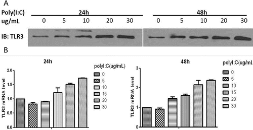
Figure 2. ELISA analysis of MUC5AC, TGF-β1, MMP9 and VEGF levels after treatment with poly(I:C). (A) The MUC5AC level increased with increasing doses of poly(I:C). (B) TGF-β1, MMP9 and VEGF expression levels increased after exposure to poly(I:C). The mean ± SD of triplicate wells from a representative experiment (n = 3) is shown.
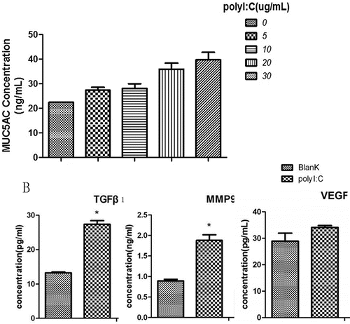
Figure 3. Western blotting analysis of p-EGFR, p-p38, p-ERK1/2 and p-JNK after exposure to poly(I:C) and of TLR3 after treatment with TLR3-NC, TLR3-siRNA1 or TLR3-siRNA2. Western blotting and RT-PCR analysis of TLR3, p-EGFR and EGFR; and ELISA analysis of MUC5AC and TGF-β1 after exposure to TLR3-siRNA2. (A) p-EGFR expression increased with increasing poly(I:C) concentration. (B) p-p38 and p-ERK1/2 were activated, whereas p-JNK was not activated. (C) Treatment with TLR3-siRNA2 for 48 hours was more effective at suppressing TLR3 than TLR3-NC or TLR3-siRNA1. (D) TLR3 production and transcription were remarkably suppressed after TLR3-siRNA2 treatment. (E) TLR3-siRNA2 decresed p-EGFR expression, but did not attenuate EGFR expression and transcription. (F) MUC5AC and TGF-β1 expression levels were diminished by treatment with TLR3-siRNA2. The mean ± SD of triplicate wells from a representative experiment (n = 3) is shown.
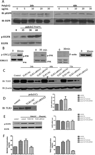
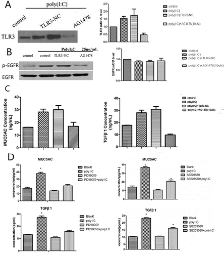
Figure 4. Western blotting and RT-PCR analysis of TLR3, p-EGFR and EGFR; ELISA analysis of MUC5AC and TGF-β1 after treatment with AG1478; and ELISA analysis of MUC5AC and TGF-β1 after pretreatment with PD98059 or SB203580. (A) TLR3 production and transcription were remarkably suppressed by AG1478. (B) Pretreatment with AG1478 decreased p-EGFR expression but did not attenuate EGFR expression and transcription. (C) MUC5AC and TGF-β1 expression levels were diminished by AG1478. (D) The poly(I:C)-induced increases in MUC5AC and TGF-β1 expression were inhibited by pretreatment with PD98059 or SB203580. The mean ± SD of triplicate wells from a representative experiment (n = 3) is shown.