6,246
Views
42
CrossRef citations to date
0
Altmetric
REVIEW ARTICLE

Desmosomes in the Heart: A Review of Clinical and Mechanistic Analyses

&
Pages 109-128 | Received 11 Mar 2014, Accepted 18 Mar 2014, Published online: 23 Apr 2014

Figures & data

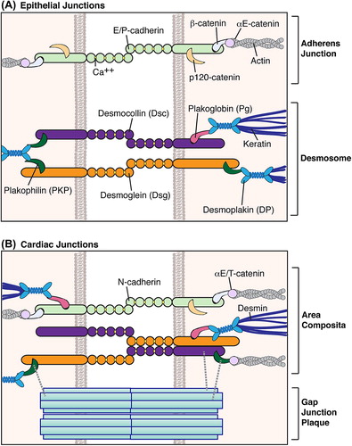
Figure 1. Intercellular junctions in epithelial and cardiac tissues. (A) In epithelial tissues, adherens junctions and desmosomes comprise discrete plaques that respectively tether actin and intermediate filaments to sites of cell–cell contact (CitationHarmon & Green, 2013; CitationSimpson et al., 2011). (B) In cardiac tissue, components of desmosomes and adherens junctions intermingle as a hybrid area composita that stabilizes adjacent gap junctions (CitationBolling & Jonkman, 2009; CitationFranke et al., 2006). Desmoplakin may localize to N-cadherin via plakoglobin (CitationCowin et al., 1986), while αT-catenin could associate with desmosomal cadherins through plakophilin 2 (CitationGoossens et al., 2007). Both desmocollin 2 and plakophilin 2 have been shown to associate with connexin43 (CitationGehmlich et al., 2011a; CitationLi et al., 2009; CitationOxford et al., 2007), potentially forming direct links between the area composita and gap junction plaques.

Figure 1. Intercellular junctions in epithelial and cardiac tissues. (A) In epithelial tissues, adherens junctions and desmosomes comprise discrete plaques that respectively tether actin and intermediate filaments to sites of cell–cell contact (CitationHarmon & Green, 2013; CitationSimpson et al., 2011). (B) In cardiac tissue, components of desmosomes and adherens junctions intermingle as a hybrid area composita that stabilizes adjacent gap junctions (CitationBolling & Jonkman, 2009; CitationFranke et al., 2006). Desmoplakin may localize to N-cadherin via plakoglobin (CitationCowin et al., 1986), while αT-catenin could associate with desmosomal cadherins through plakophilin 2 (CitationGoossens et al., 2007). Both desmocollin 2 and plakophilin 2 have been shown to associate with connexin43 (CitationGehmlich et al., 2011a; CitationLi et al., 2009; CitationOxford et al., 2007), potentially forming direct links between the area composita and gap junction plaques.

Table 1. Cardiomyopathy (CM) classifications. Generalized clinical findings and etiology of each condition, in addition to those of unclassified cardiomyopathies (left ventricular noncompaction, stress-induced, cirrhotic, etc.) are further described in referenced reviews (CitationElliott, 2008; CitationHarvey & Leinwand, 2011; CitationMaron et al., 2006).

Table 2. Mutations associated with AC and AC-overlap syndromes. Detailed descriptions of the listed mutations can be found in the literature cited in this text and elsewhere (CitationRickelt & Pieperhoff, 2012; Citationvan der Zwaag et al., 2009).

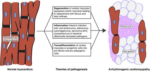
Figure 2. Theories of AC pathogenesis. AC may arise from cardiac myocyte degeneration, inflammation of myocardium from infection by cardiotropic pathogens, and/or transdifferentiation of cardiac myocytes or progenitor cells into fat- and fibrotic scar-producing cells (CitationBasso et al., 2011; CitationHerren et al., 2009).

Figure 2. Theories of AC pathogenesis. AC may arise from cardiac myocyte degeneration, inflammation of myocardium from infection by cardiotropic pathogens, and/or transdifferentiation of cardiac myocytes or progenitor cells into fat- and fibrotic scar-producing cells (CitationBasso et al., 2011; CitationHerren et al., 2009).

Table 3. Potential mechanisms of AC mutation pathogenicity. Select findings from mouse models and/or in vitro studies are highlighted here, with further information available in the text.
Reprints and Corporate Permissions

Please note: Selecting permissions does not provide access to the full text of the article, please see our help page How do I view content?

To request a reprint or corporate permissions for this article, please click on the relevant link below:

Academic Permissions

Please note: Selecting permissions does not provide access to the full text of the article, please see our help page How do I view content?

Obtain permissions instantly via Rightslink by clicking on the button below:

If you are unable to obtain permissions via Rightslink, please complete and submit this Permissions form. For more information, please visit our Permissions help page.