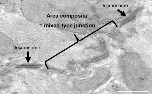
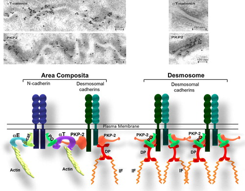
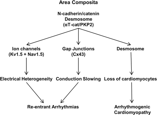
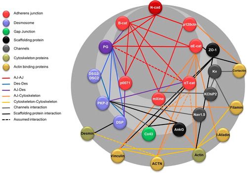
2,181
Views
44
CrossRef citations to date
0
Altmetric
Review Article

N-Cadherin/Catenin Complex as a Master Regulator of Intercalated Disc Function

&
Pages 169-179 | Received 20 Mar 2014, Accepted 24 Mar 2014, Published online: 28 Apr 2014

Figures & data

Figure 1. Transmission electron microscopy of a cross section through intercalated disc of a mouse heart. TEM image showing the intercalated disc with a large mixed-type junction (area composita) surrounded by two dense desmosome structures (black arrows).

Figure 1. Transmission electron microscopy of a cross section through intercalated disc of a mouse heart. TEM image showing the intercalated disc with a large mixed-type junction (area composita) surrounded by two dense desmosome structures (black arrows).

Figure 2. Molecular organization of area composita vs. desmosome in the heart. (a) Immunoelectron microscopic images of the myocardium of a mouse heart showing the co-localization of PKP2 and αT-catenin at the area composita. Note the absence of αT-catenin in the desmosome. These images were originally published in a study by CitationGoossens et al. (2007). (b) Model for cadherin-based cell–cell adhesion in the heart. This model represents the composition of a hybrid junction compared to a desmosome. αT-Catenin recruits desmosomal proteins through its interaction with plakophilin-2 (PKP2), forming a hybrid junction (named area composita), reinforcing the intercalated discs resistance. (αE: αE-catenin; αT: αT-catenin; β-cat: β-catenin; PG: plakoglobin; PKP2: plakophillin-2; IF: intermediate filaments).

Figure 2. Molecular organization of area composita vs. desmosome in the heart. (a) Immunoelectron microscopic images of the myocardium of a mouse heart showing the co-localization of PKP2 and αT-catenin at the area composita. Note the absence of αT-catenin in the desmosome. These images were originally published in a study by CitationGoossens et al. (2007). (b) Model for cadherin-based cell–cell adhesion in the heart. This model represents the composition of a hybrid junction compared to a desmosome. αT-Catenin recruits desmosomal proteins through its interaction with plakophilin-2 (PKP2), forming a hybrid junction (named area composita), reinforcing the intercalated discs resistance. (αE: αE-catenin; αT: αT-catenin; β-cat: β-catenin; PG: plakoglobin; PKP2: plakophillin-2; IF: intermediate filaments).

Table 1. Genetic manipulation of adherens junction proteins in the adult myocardium.

Figure 3. Functional hierarchy at the intercalated disc. Scheme depicts the relationship between the different junctions and cardiac dysfunction.

Figure 3. Functional hierarchy at the intercalated disc. Scheme depicts the relationship between the different junctions and cardiac dysfunction.

Figure 4. Junctional proteins interactome. The intercalated disc protein–protein interaction network is based on STRING v9.1 analysis (http://www.string-db.org/) using the default settings (CitationFranceschini et al., 2013). Black lines represent direct interactions, while dotted lines represent assumed interactions based on the studies of the structures homology. (Ncad: N-cadherin; B-cat: β-catenin; p120ctn: p120-catenin; αE-cat: αE-catenin; αT-cat: αT-catenin; PG: plakoglobin; PKP2: plakophillin-2; DSG2: desmoglein-2; DSC2: desmocollin-2; DSP: desmoplakin; Cx43: connexin-43; AnkG: ankyrin G; Kv: potassium channels; kChiP2: Kv channel-interacting protein 2; ACTN: actinin).

Figure 4. Junctional proteins interactome. The intercalated disc protein–protein interaction network is based on STRING v9.1 analysis (http://www.string-db.org/) using the default settings (CitationFranceschini et al., 2013). Black lines represent direct interactions, while dotted lines represent assumed interactions based on the studies of the structures homology. (Ncad: N-cadherin; B-cat: β-catenin; p120ctn: p120-catenin; αE-cat: αE-catenin; αT-cat: αT-catenin; PG: plakoglobin; PKP2: plakophillin-2; DSG2: desmoglein-2; DSC2: desmocollin-2; DSP: desmoplakin; Cx43: connexin-43; AnkG: ankyrin G; Kv: potassium channels; kChiP2: Kv channel-interacting protein 2; ACTN: actinin).

Reprints and Corporate Permissions

Please note: Selecting permissions does not provide access to the full text of the article, please see our help page How do I view content?

To request a reprint or corporate permissions for this article, please click on the relevant link below:

Academic Permissions

Please note: Selecting permissions does not provide access to the full text of the article, please see our help page How do I view content?

Obtain permissions instantly via Rightslink by clicking on the button below:

If you are unable to obtain permissions via Rightslink, please complete and submit this Permissions form. For more information, please visit our Permissions help page.