Figures & data
Table 1. Effect of 24 h, 48 h, and 6 day TBBPA or 1 h followed by 24-h, 48-h, or 6-day periods in TBBPA-free media on human NK cell viability.
Figure 1. Effects of 24-h TBBPA exposures on NK cell-surface protein expression. Values are the mean (± SD) of mean fluorescence intensity (MFI) combined from four different experiments each using a different donor (n = 4). Control cells were vehicle treated. *Statistically significant decrease (p < 0.05) analyzed as described in the Materials and methods. NK, natural killer; TBBPA, tetrabromobisphenol A.
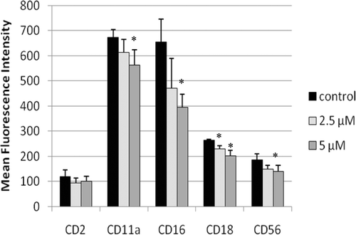
Figure 2. Representative histograms from studies of effects of 24-h exposures to 5 µM TBBPA on NK cell-surface protein expression. (A) CD11a (control mean fluorescence intensity (MFI) = 643.32, TBBPA MFI = 512.52); (B) CD16 (control MFI = 580.52, TBBPA MFI = 390.5); (C) CD18 (control MFI = 259.63, TBBPA MFI = 210.54); and (D) CD56 (control MFI = 68.54, TBBPA MFI = 46.54). Dotted line: IgG control; thin solid line: control NK cells + appropriate antibody; bold line: TBBPA-exposed cells + appropriate antibody. Y-axis: cell number; X-axis: fluorescence intensity. (E) Dot-plot of cell preparation used in experiment. Shifts in fluorescence intensity were essentially the same whether gated (R1) or un-gated cell populations were used. NK, natural killer; TBBPA, tetrabromobisphenol A.
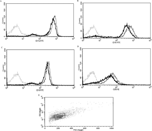
Figure 3. Effects of 48-h TBBPA exposures on NK cell-surface protein expression. Values are the mean ± S.D. of mean fluorescence intensity (MFI) combined from four different experiments each using a different donor (n = 4). *Statistically significant decrease (p < 0.05). NK, natural killer; TBBPA, tetrabromobisphenol A.
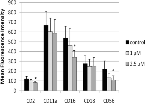
Figure 4. Representative histograms from studies of effects of 48-h exposures to 2.5 µM TBBPA on NK cell-surface protein expression. (A) CD2 (control MFI = 123.71, TBBPA MFI = 79.36); (B) CD16 (control MFI = 678.42, TBBPA MFI = 442.22); and (C) CD56 (control MFI = 277.39, TBBPA MFI = 154.74). Dotted line: IgG control; thin solid line: control NK cells + appropriate antibody; bold line: TBBPA-exposed cells + appropriate antibody. Y-axis: cell number; X-axis: fluorescence intensity. (D) Dot-plot of cell preparation used in experiment. NK, natural killer; TBBPA, tetrabromobisphenol A.
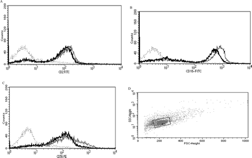
Figure 5. Effects of 6-day TBBPA exposures on NK cell-surface protein expression. Values shown are the mean ± SD of mean fluorescence intensity combined from four different experiments each using a different donor (n = 4). *Statistically significant decrease (p < 0.05). NK, natural killer; TBBPA, tetrabromobisphenol A.
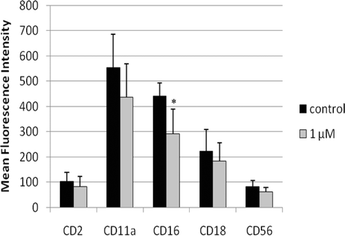
Figure 6. Representative histograms from studies of effects of 6-day exposures to 2.5 µM TBBPA on CD16 expression in NK cells. (A) CD16 (control MFI = 392.05, TBBPA MFI = 182.58). Dotted line: IgG control; thin solid line: control NK cells + appropriate antibody; bold line: TBBPA-exposed cells + appropriate antibody. Y-axis: cell number; X-axis: fluorescence intensity. (B) Dot-plot of cell preparation used in experiment. NK, natural killer; TBBPA, tetrabromobisphenol A.
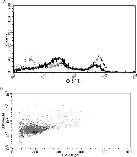
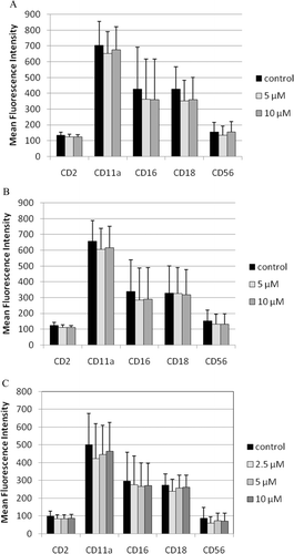
Figure 7. Effects of 1-h exposures to TBBPA—followed by 24-h, 48-h, or 6-day incubations in TBBPA-free media—on NK cell-surface protein expression. (A) One-hour exposures to 10 µM and 5 µM TBBPA followed by 24-h incubation in TBBPA-free media. (B) One-hour exposures to 10 µM and 5 µM TBBPA followed by 48-h incubation in TBBPA-free media. (C) One-hour exposures to 10 µM, 5 µM, and 2.5 µM TBBPA followed by 6-day incubation in TBBPA-free media. Values shown are the mean ± SD of mean fluorescence intensity (MFI) combined from three different experiments each using a different donor (n = 3). NK, natural killer; TBBPA, tetrabromobisphenol A.