Figures & data
Table 1. Percentage of AFM1 removal from PBS and reconstituted milk by Lactobacillus plantarum MON03 and Lactobacillus rhamnosus GAF01 strains.
Figure 1. High performance liquid chromatography (HPLC) chromatograms. (a) Untreated (positive control) aflatoxin M1 (at 0.15 µg AFM1/ml)-phosphate buffered saline (PBS) solution; (b) AFM1-free PBS solution; or (c) AFM1 (0.15 µg/ml)-PBS solution after 6 h treatment with Lactobacillus rhamnosus GAF01 at 108 CFU/ml.
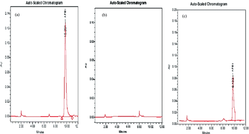
Figure 2. Effect of washing on bacteria:AFM1 complex. Initial binding was determined after bacteria (108 CFU/ml) and AFM1 (0.05 µg/ml) were incubated together for 6 h at 37°C. Bacteria/AFM1 complexes formed were subjected to centrifugation and then washing with 5 ml PBS, before the amount of AFM1 released was determined as outlined in the Methods. Results shown are the mean (± SD) from triplicate samples.
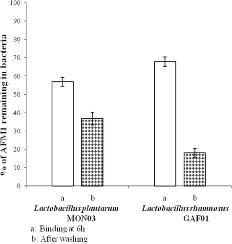
Figure 3. Influence of AFM1 and Lactobacillus rhamnosus GAF01 (alone or in combination) on lymphocyte sub-types. Balb/c mice received daily (for 14 days) by gavage distilled water (open bar), AFM1 (100 µg/kg BW) (solid bar), L. rhamnosus GAF01 (1 mg/kg BW) (shaded bold bar), or AFM1 + L. rhamnosus GAF01 (shaded fine bar). Lymphocyte counts were then performed on blood samples collected on Day 15 of the experiment (i.e., 1 day after final exposure in each regimen). Results are expressed as mean [± SE] from n = 12 mice/group. A Student’s t-test was used to compare differences between groups: aValue significantly/bnot significantly different from control at p ≤ 0.05.
![Figure 3. Influence of AFM1 and Lactobacillus rhamnosus GAF01 (alone or in combination) on lymphocyte sub-types. Balb/c mice received daily (for 14 days) by gavage distilled water (open bar), AFM1 (100 µg/kg BW) (solid bar), L. rhamnosus GAF01 (1 mg/kg BW) (shaded bold bar), or AFM1 + L. rhamnosus GAF01 (shaded fine bar). Lymphocyte counts were then performed on blood samples collected on Day 15 of the experiment (i.e., 1 day after final exposure in each regimen). Results are expressed as mean [± SE] from n = 12 mice/group. A Student’s t-test was used to compare differences between groups: aValue significantly/bnot significantly different from control at p ≤ 0.05.](/cms/asset/0eea1276-3d0a-4afc-bdab-861daf208dec/iimt_a_718810_f0003_b.gif)
Figure 4. Influence of AFM1 and Lactobacillus rhamnosus GAF01—alone or in combination—on blood cell counts. Balb/c mice received daily (for 14 days) by gavage distilled water (open bar), AFM1 (100 µg/kg BW) (solid bar), L. rhamnosus GAF01 (1 mg/kg BW) (shaded bold bar), or AFM1 + L. rhamnosus GAF01 (shaded fine bar). Erythrocyte and leukocyte counts were then performed on blood samples collected on Day 15 of the experiment (i.e., 1 day after final exposure in each regimen). Results are expressed as mean [± SE] from n = 12 mice/group. A Student’s t-test was used to compare differences between groups: aValue significantly/bnot significantly different from control at p ≤ 0.05.
![Figure 4. Influence of AFM1 and Lactobacillus rhamnosus GAF01—alone or in combination—on blood cell counts. Balb/c mice received daily (for 14 days) by gavage distilled water (open bar), AFM1 (100 µg/kg BW) (solid bar), L. rhamnosus GAF01 (1 mg/kg BW) (shaded bold bar), or AFM1 + L. rhamnosus GAF01 (shaded fine bar). Erythrocyte and leukocyte counts were then performed on blood samples collected on Day 15 of the experiment (i.e., 1 day after final exposure in each regimen). Results are expressed as mean [± SE] from n = 12 mice/group. A Student’s t-test was used to compare differences between groups: aValue significantly/bnot significantly different from control at p ≤ 0.05.](/cms/asset/33a41ebe-564c-491a-a5d4-1f8409b3cd09/iimt_a_718810_f0004_b.gif)