Figures & data
Figure 1. Anakinra blocks IL-1α from pig origin in a cell-based bio-assay. D10 cells were cultured with varying concentrations of rpigIL-1α (A) or with 3 ng/ml rpigIL-1α plus varying concentrations of anakinra (B). After a 4-day incubation, MTT was added to the cultures for 3 h followed by 10% SDS/50% DMF. Absorbance in each well was then measured at 590 nm. For each treatment group, mean (± SE) is depicted. O = concentration rpigIL-1α used in (B).
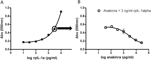
Table 1. Mean relative organ weights and terminal body weights following anakinra treatment.
Figure 2. Serum levels of anti-anakinra antibodies. Blood for serum preparation was taken on several study days prior to treatment and analyzed for antibodies to anakinra. As the concentrations are calculated based on a standard curve using a goat anti-human IL-1ra antibody, depicted concentrations of pig-anti-anakinra antibodies should be considered estimated concentrations. (A) For each treatment group, mean ± SEM is depicted. -o-, Placebo control females; -•-, Placebo control males; -□-, Low dose anakinra females; -▪-, Low dose anakinra males; -◊-, High dose anakinra females; -▪-, High dose anakinra males. Day 7: 2-way ANOVA, placebo control vs low dose, p < 0.05. Furthermore, no significant differences are observed between groups. (B) Individual data anti-anakinra antibodies on study day 30.
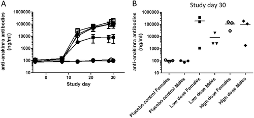
Table 2. Pharmacokinetic parameters at Day 0 and Day 28 for anakinra following daily dosing at 0.5 and 5.0 mg/kg body weight to male and female Göttingen minipigs.
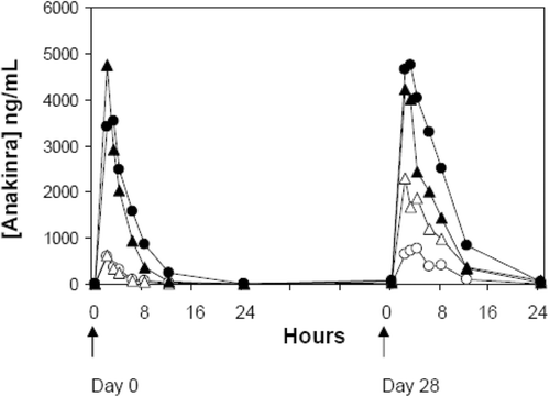
Figure 3. Anakinra plasma concentration vs time curves. Data shown are from Göttingen minipigs following a subcutaneous low (-o-) and high (-•-) dose (females) and low (-Δ-) and high (-▴-) dose (males) at Day 0 and Day 28. Mean values for each group are depicted.