Figures & data
Figure 1. Chemical structures of the four tested aflatoxins (AF). Note that AFB2 and AFG2 are derivatives of AFB1 and AFG1, respectively. OMe = methoxy.
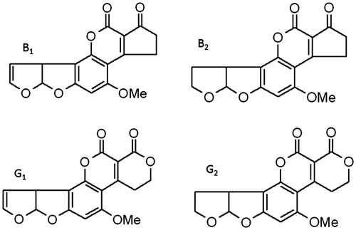
Figure 2. Effect of naturally occurring levels of AF on phagocytic activity of porcine monocyte-derived dendritic cells (MoDC). MoDC isolated from each of three piglets were separately treated for different periods of time with a mixture of several aflatoxins (mAF) containing AFB1 (2.0 ng/ml), AFB2 (1.0 ng/ml), AFG1 (0.5 ng/ml), and AFG2 (0.5 ng/ml). At the end of each period, 5.0 × 10⁶ fluorescent 1.0-μm microparticles were added to each set of MoDC to assess if there were effects on phagocytic activity induced by the treatments. Data are presented as mean (±SEM, n = 3) percent (%) change in phagocytic activity relative to values for each piglet’s corresponding untreated MoDC. Dashed line = phagocytic activity of control non-AF-treated MoDC.
Figure 3. Aflatoxins affect the cell surface expression of DC activation markers. MoDC isolated from each of three piglets were separately treated with a mixture of several aflatoxins (mAF containing AFB1 [2.0 ng/ml], AFB2 [1.0 ng/ml], AFG1 [0.5 ng/ml], and AFG2 [0.5 ng/ml]) for 12 and 24 h and then analyzed for expression of CD40, CD80/86, CD25, and MHC-II by flow cytometry. Data are presented as mean (±SEM, n = 3) percent (%) change in expression relative to values for each piglet’s corresponding untreated MoDC. *p < 0.05 to untreated cells. imm = immature (untreated or control) MoDC.
![Figure 3. Aflatoxins affect the cell surface expression of DC activation markers. MoDC isolated from each of three piglets were separately treated with a mixture of several aflatoxins (mAF containing AFB1 [2.0 ng/ml], AFB2 [1.0 ng/ml], AFG1 [0.5 ng/ml], and AFG2 [0.5 ng/ml]) for 12 and 24 h and then analyzed for expression of CD40, CD80/86, CD25, and MHC-II by flow cytometry. Data are presented as mean (±SEM, n = 3) percent (%) change in expression relative to values for each piglet’s corresponding untreated MoDC. *p < 0.05 to untreated cells. imm = immature (untreated or control) MoDC.](/cms/asset/bb2849d2-897b-47ad-8523-f8752cde8b39/iimt_a_916366_f0003_c.jpg)
Figure 4. Aflatoxins affect the T-cell stimulatory capacity of porcine MoDC. MoDC isolated from each of three piglets were separately treated with a mixture of several aflatoxins (see legend) for 12 and 24 h. The mAF-treated MoDC were subsequently co-cultured with 2.0 × 10⁵ CD6+ T-cells at a 1:30 (DC:T) ratio. T-cell proliferation was measured via [3H]-methyl thymidine incorporation. Data are presented as mean (±SEM, n = 3) stimulation index of the three individual sets of piglet cells analyzed. *p < 0.05 vs non-AF-treated MoDC.
![Figure 4. Aflatoxins affect the T-cell stimulatory capacity of porcine MoDC. MoDC isolated from each of three piglets were separately treated with a mixture of several aflatoxins (see Figure 3 legend) for 12 and 24 h. The mAF-treated MoDC were subsequently co-cultured with 2.0 × 10⁵ CD6+ T-cells at a 1:30 (DC:T) ratio. T-cell proliferation was measured via [3H]-methyl thymidine incorporation. Data are presented as mean (±SEM, n = 3) stimulation index of the three individual sets of piglet cells analyzed. *p < 0.05 vs non-AF-treated MoDC.](/cms/asset/96c54172-1cdf-40e4-9610-3a38d00a5996/iimt_a_916366_f0004_c.jpg)
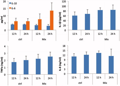
Figure 5. Cytokine expression pattern of AF-exposed MoDC. MoDC isolated from each of three piglets were separately treated with either 0 ng/ml (ctrl) or a mixture of aflatoxins (see legend) for 12 and 24 h. Cell culture supernatants harvested at the end of each incubation period were analyzed for the presence of IL-1β, IL-6, IL-8, IL-10, and TNFα by ELISA. Data are presented as mean (±SEM, n = 3) levels of each cytokine in the medium (at each timepoint) from the three individual sets of piglet cells analyzed. Ctrl = non-AF-treated MoDC.