Figures & data
Figure 1. Transmission electronic microscopy (TEM) image of primary AgNP70 stock solution and evaluation of cell viability/morphology in AgNP70-induced human PMN. (A) Samples of the manufacturer stock solution was used for characterization by TEM to confirm the primary diameter of the NP was ∼70 nm. Freshly isolated human PMN were incubated for 24 h in the presence of buffer (controls), 10 or 100 μg/ml AgNP70, 65 ng/ml GM-CSF (GM) or 100 μg/ml AgNP20 and then (B) cell necrosis was evaluated by measuring levels of LDH released into the culture supernatants, (C) number of cells remaining in plates were determined and (D) cell morphology was evaluated using optical microscopy (400×). Results shown are means ± SEM (n = 3 [B]; n = 5 [C]). Both low and high controls were used according to manufacturer recommendations. AgNP20 = silver nanoparticles of 20 nm; arrowhead = aggregates; arrows = increased cell size in AgNP20-induced PMN. Pictures in (D) are from one representative experiment out of five done with different blood donors.
![Figure 1. Transmission electronic microscopy (TEM) image of primary AgNP70 stock solution and evaluation of cell viability/morphology in AgNP70-induced human PMN. (A) Samples of the manufacturer stock solution was used for characterization by TEM to confirm the primary diameter of the NP was ∼70 nm. Freshly isolated human PMN were incubated for 24 h in the presence of buffer (controls), 10 or 100 μg/ml AgNP70, 65 ng/ml GM-CSF (GM) or 100 μg/ml AgNP20 and then (B) cell necrosis was evaluated by measuring levels of LDH released into the culture supernatants, (C) number of cells remaining in plates were determined and (D) cell morphology was evaluated using optical microscopy (400×). Results shown are means ± SEM (n = 3 [B]; n = 5 [C]). Both low and high controls were used according to manufacturer recommendations. AgNP20 = silver nanoparticles of 20 nm; arrowhead = aggregates; arrows = increased cell size in AgNP20-induced PMN. Pictures in (D) are from one representative experiment out of five done with different blood donors.](/cms/asset/61c2a9ac-bbbc-4d83-8990-79dc3730704b/iimt_a_1106622_f0001_c.jpg)
Table 1. Size and Zeta potential of AgNP20 and AgNP70.
Figure 2. AgNP70 and AgNP20 have opposing effect on human neutrophil apoptosis, but both act as potent inhibitors of de novo protein synthesis without inducing ROS production. Freshly isolated human PMN were incubated with the indicated agonist for the specified periods of time and apoptosis (A) and then de novo protein synthesis (B) and ROS production (C) were determined (see Materials and methods). Results shown are means ± SEM (n = 4); (C) Results are from one representative experiment out of four performed with different blood donors. VAA, Viscum album agglutinin-I (1 μg/ml); PMA, phorbol-12-myristate-13-acetate (10−7 M); MW, molecular weights; CHX, cycloheximide (10 μg/ml); and ATO, arsenic trioxide (5 μM).
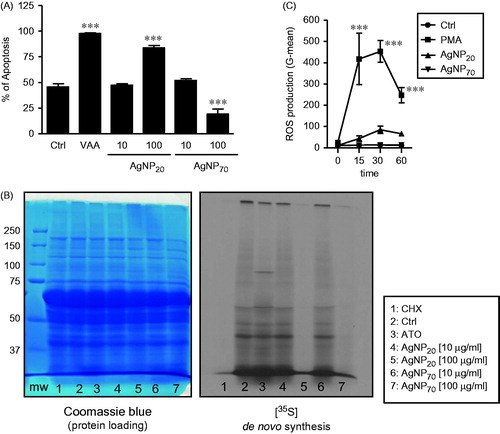
Figure 3. AgNP20 promote release of IL-8 at a concentration that does not induce neutrophil apoptosis when exogenously added to the cultures. (A) Freshly isolated human PMN were incubated for 24 h in the presence of buffer (control/Crtl), 65 ng/ml GM-SCF (GM), 1 μg/ml VAA-I (VAA), 10 or 100 μg/ml of AgNP70 or AgNP20 and the release of IL-8 into extracellular milieu was then quantified by ELISA. (B) Cells were incubated for 24 h in the presence of 0 (Ctrl), 10, 25, 50 or 100 ng/ml recombinant human IL-8 and apoptosis then determined by cytology (see Materials and methods). Results shown are means ± SEM (n = 3/treatment).
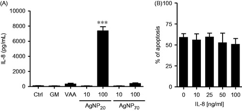
Figure 4. AgNP20 but not AgNP70 induce degranulation and release of albumin and MMP-9. Freshly isolated human PMN were incubated for 30 or 60 min with buffer (Ctrl), 10−8 M fMLF, 100 μg/ml AgNP20 or 100 μg/ml AgNP70 and the release of (A) albumin and (B) MMP-9 into the extracellular milieu was then detected by Western blot. Densitometry analysis (right part of each panel) was performed and results (means ± SEM, n ≥ 3/treatment) are expressed in arbitrary units derived from integrated density values (IDV). Left part: one representative experiments out of three (A) or four (B).
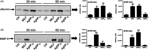
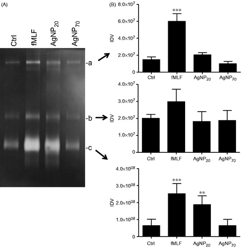
Figure 5. AgNP20 but not AgNP70 increase gelatinolytic activity in human PMN. Freshly isolated human PMN were isolated and incubated with the buffer (Ctrl), fMLF (10−8 M) or with 100 μg/ml of AgNP20 or AgNP70 for 60 min and the gelatinolytic activity of MMP-9 was then assessed by zymography. (A) Typical results where the enzymatic activity was observed by appearance of clear bands (a, b and c). (B) Densitometric analysis of corresponding bands, expressed as integrated density values (IDV). Results shown are means ± SEM (n = 3–5/treatment).