
Figures & data
Figure 1. Effects of exposures to TBBPA on IL-1β secretion from human NK cells, monocyte-depleted PBMC and PBMC. (a) NK cells (donor KB129). (b) Monocyte-depleted PBMC (donor F177). (c) PBMC (donor F188). All doses 0.05–5.0 μM TBBPA. Data shown are means ± SD. * Value significantly different from appropriate control (0 μM) at p < 0.05.
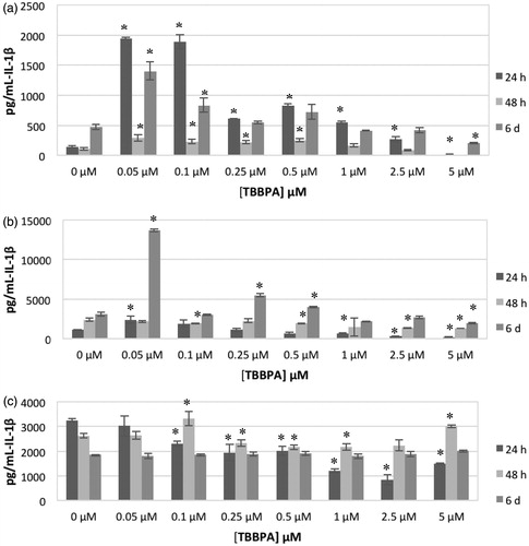
Table 1. Effects of TBBPA exposures on IL-1β secretion from highly purified human NK cells.
Table 2. Effects of TBBPA exposures to on IL -1β secretion from monocyte-depleted PBMC.
Table 3. Effects of TBBPA exposures to TBBPA on IL-1β secretion from PBMC.
Table 4. Effects of HBCD exposures to on IL -1β secretion from highly-purified human NK cells.
Figure 2. Effects of exposures to HBCD on IL-1β secretion from human NK cells, monocyte-depleted PBMC and PBMC. (a) NK cells (donor KB181). (b) Monocyte-depleted PBMC (donor F202). (c) PBMC (donor F189). All doses 0.05–5.0 μM HBCD. Data shown are means ± SD. * Value significantly different from appropriate control at p < 0.05.
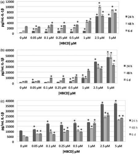
Table 5. Effects of HBCD exposures on IL -1β secretion from monocyte-depleted PBMC.
Table 6. Effects of HBCD exposures on IL -1β secretion from PBMC.
Figure 3. Effects of 24-h exposure to HBCD on IL-1β secretion from monocyte-depleted PBMC pre-treated with selective enzyme inhibitors. Data shown is based on cells from an individual donor. (a) Caspase I inhibitor II (Caspase I) (donor F 267). (b) NF-κB inhibitor (BAY 11-7085) (donor F275). (c) JNK inhibitor (donor F271). (d) MEK1/2 (ERK1/2 pathway) inhibitor (PD98059) (donor F267). (e) p38 inhibitor (SB202190) (donor F255). * Value significantly different from control cells with vehicle alone at p < 0.05. ** Value significantly different from control + inhibitor at p < 0.05.
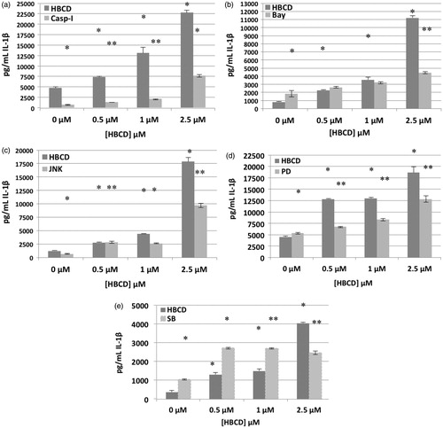
Figure 4. Signaling cascade involved in IL-1β secretion.

Table 7. Effects of 24 h exposure to HBCD ± pathway inhibitors on IL-1β secretion from MD-PBMC.
Table 8. Effects of 24 h exposure to HBCD ± pathway inhibitors on IL-1β secretion from MD-PBMC.
Table 9. Effects of 24 h exposure to HBCD ± pathway inhibitors on IL-1β secretion from MD-PBMC.
Table 10. Effects of 24 h exposure to HBCD ± pathway inhibitors on IL-1β secretion from MD-PBMC.
Table 11. Effects of 24 h exposure to HBCD ± pathway inhibitors on IL-1β secretion from MD-PBMC.