
Figures & data
Figure 1. Composite macroscopic views of body and genito-urinary system: A. Frontal view shows small and anteverted nose, contracted elbows, and broad hands. The vertical, midline scar extending from chest to abdomen is secondary to aortopexy. There is a gastrostomy tube in place. B. Shows distended abdomen, hypoplastic chest, undescended testes, and bilateral talus deformity. C. Dissected kidneys, ureters, and urinary bladder in anatomic position. Note multicystic and dysplastic kidney with intermixed diffuse nodular parenchyma. The urinary bladder is enlarged as seen in obstructive renal dysplasia; however, the ureters are not distended. D. Posterior view of longitudinal section of the kidneys shows scattered multicystic renal parenchyma with lack of normal cortico-medullary demarcation, mixed with solid, nodular areas.
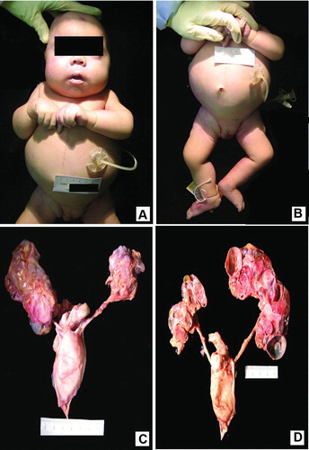
Figure 2. Composite photomicrograph of renal parenchyma: A. Hematoxylin-eosin [H&E] stain with 40 X-lens shows clusters of highly cellular sheets of primitive small blue cells with scant cytoplasm, representing the blastema component of NB that is surrounded by few epithelial tubules and fibrous stroma. B. Immunohistochemistry stain using anti PAX2 antibody (20-X lens) is immune-positive in the areas of primitive renal blastema with glomeruloid features. C. Representative immunohistochemistry image using anti WT-1 antibody. (20 X-lens). The areas of NB are strongly immunoreactive for WT-1. D. Immunostain using anti GLEPP-1 (glomerular epithelial protein) antibody (20 X-lens). Note that mature renal glomeruli stained positive, while the blastema component is negative. 103 × 77 mm (300 × 300 DPI).
![Figure 2. Composite photomicrograph of renal parenchyma: A. Hematoxylin-eosin [H&E] stain with 40 X-lens shows clusters of highly cellular sheets of primitive small blue cells with scant cytoplasm, representing the blastema component of NB that is surrounded by few epithelial tubules and fibrous stroma. B. Immunohistochemistry stain using anti PAX2 antibody (20-X lens) is immune-positive in the areas of primitive renal blastema with glomeruloid features. C. Representative immunohistochemistry image using anti WT-1 antibody. (20 X-lens). The areas of NB are strongly immunoreactive for WT-1. D. Immunostain using anti GLEPP-1 (glomerular epithelial protein) antibody (20 X-lens). Note that mature renal glomeruli stained positive, while the blastema component is negative. 103 × 77 mm (300 × 300 DPI).](/cms/asset/b6eca4bc-9756-454e-ac69-775f48c1d9c3/ipdp_a_1014952_f0002_c.jpg)