Figures & data
Table 1. Maternal and fetal information for our four patients.
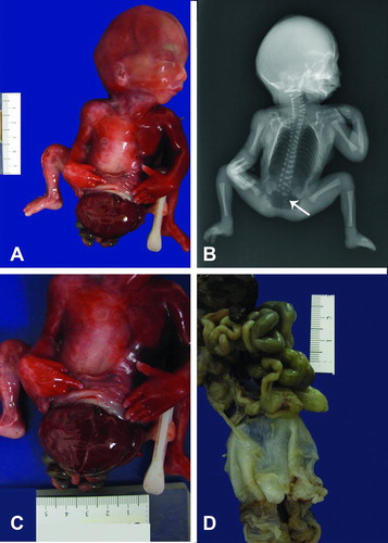
Figure 1. (A) Posterior view of Patient 1 showing fetus with sacrolumbar myelomeningocele, and contracted lower extremities. (B) Lateral fetal view depicts the attached amnion covering omphalocele with a short umbilical cord wrapped around the amnion covering the abdominal wall defect. The fetus also has bilateral flexion contracture deformity of lower extremities. (C) Post-mortem antero-posterior x-ray showing segmentation anomalies of the thoracic vertebrae (thick arrow), omphalocele (thin arrow) and meconium peritonitis (small thin arrows). (D) Post-mortem lateral x-ray showing lumbosacral myelomeningocele (arrow).
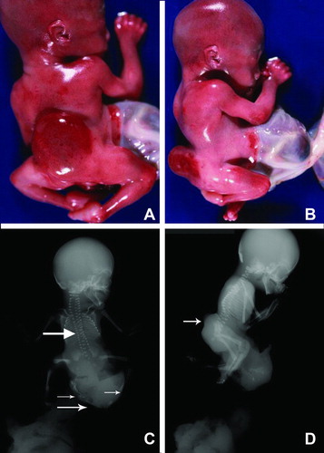
Figure 2. (A) Gross picture of Patient 1 showing fetus with attached cord and placenta. (B) Closer view of short umbilical cord covered with amnion. Notice a large interface of abdominal skin with omphalocele sac.
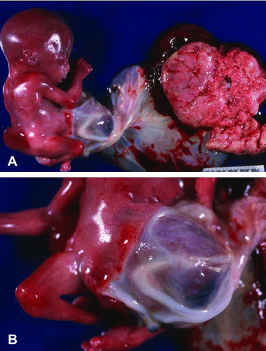
Figure 3. Gross photograph of Patient 2 showing fetus with a right antero-lateral body wall thoracoabdominoschisis covered by amnion. The right upper extremity is missing, the umbilical cord is short and the lower extremities are slightly contracted.
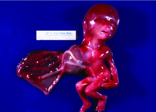
Figure 4. (A–B). Gross picture of macerated fetus after formalin fixation showing club-feet deformity and an abdominal wall defect. The lower extremities are contracted. (C–D) Post-mortem antero-posterior and lateral x-rays showing severe levoscoliosis, widely separated ischial ossification centers (larger arrows), and external protrusion of abdominal contents (smaller arrow), and club foot deformity.
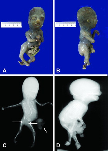
Figure 5. (A) Gross photograph of fetus with no external genitalia and omphalocele sac involving abdomen and pelvis. Notice flexion deformity of both lower extremities. (B) Post-mortem antero-posterior x-ray showing a large lucency occupuing the chest and abdomen consistent with post-mortem air, there are vertebral body segmentation anomalies and hypoplastic sacrum (arrow). (C) Photograph depicts a closer view of extracorporeal organs. (D) Gross picture of formalin fixed organ block of Patient 4. The lower portion demonstrates the omphalocele sac still attached to abdomino-pelvic organs.