Figures & data
Fig. 1. Electrocardiogram (ECG) showing sinus tachycardia with poor R-wave progression in precordial leads.
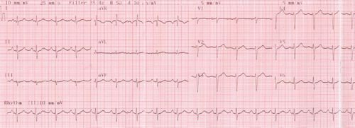
Fig. 2. (A and B) Selective left coronary angiogram showing severe thrombotic stenosis at the proximal portion of the left anterior descending artery (yellow arrows). (C and D) Selective left coronary angiogram obtained 3 days after intravenous Tirofiban infusion showing patent left anterior descending artery without residual stenosis.
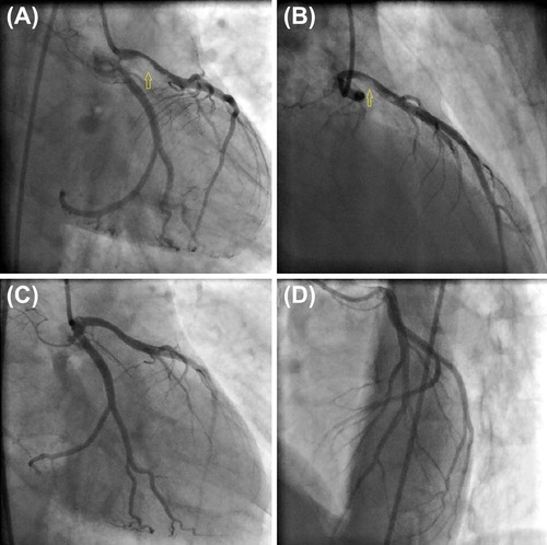
Table 1. Case reports of acute myocardial infarction associated with thinner abuse.
Supplemental material