Figures & data
Table 1. Toxicity of Ag, ZnO and CuO nanoparticles (NPs) and their respective ions to bacteria, yeast, algae, crustaceans, fish and mammalian cells in vitro.
Table 2. Classification of journals in which toxicity data for Ag, ZnO or CuO NPs and different test organisms or cells were published and that are cited in the current Review.
Figure 1. Number and share of papers in ISI WoS on May 5th and 6th, 2013 concerning uptake, oxidative stress and other mechanistic nanotoxicological information (damage to membranes, mitochondria, DNA and lipid peroxidation) on Ag (A), ZnO (B) and CuO NPs (C) for bacteria, yeast, algae, daphnids, fish and mammalian cells in vitro separately and for all these organism groups (TOTAL). Data are plotted from Supplemental Table SIII.
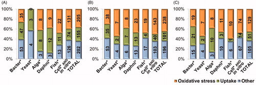
Figure 2. Uptake and attachment of NPs on cellular membranes. (A) PVP coated Ag NPs interacting with the membrane of bacteria Escherichia coli (TEM image), (B) CuO NPs in the close vicinity of the cell wall of yeast Saccharomyces cerevisiae (dyed with trypan blue, light microscopy image), (C) Ag NPs taken up by protozoa Tetrahymena thermophila into its food vacuoles (light microscopy image), (D) Light microscopy image of Daphnia magna exposed to CuO NPs: Ag NPs inside the gut and interacting with the gut epithelium (cross-section).
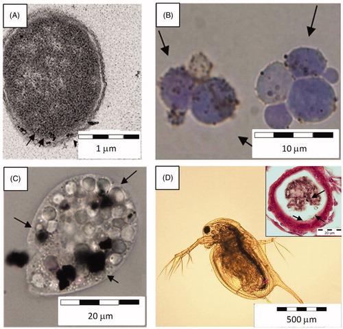
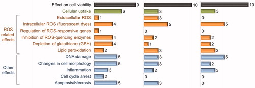
Figure 3. Summary on adverse effects of Ag, CuO and ZnO nanoparticles in mammalian cell cultures. Ten papers were selected for each nanoparticle; numbers on graphs show the number of articles where the indicated effect was studied and observed as a positive result (data are summarized from Supplemental Tables SIV–SVI).