Figures & data
Figure 1. AP illustration of the scapula showing the 14 coracoid fracture patterns seen in this cohort. The patterns together yield the “coracoid fracture map.”
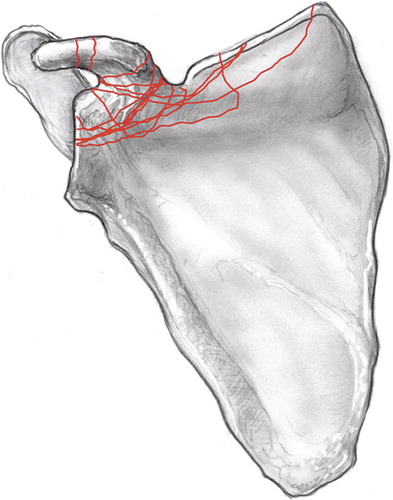
Figure 2. AP illustration of the scapula showing the 13 acromion fracture patterns seen in this cohort. The patterns together yield the “acromion fracture map.”
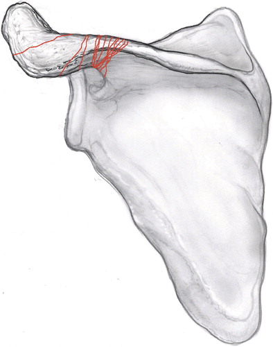
Figure 3 A. 2D- and 3D-CT scan of the scapula (patient 9) showing a minimally displaced fracture of the acromial base at the time of injury. B. AP radiography (left) and 2D-CT (right) of the scapula in the same patient 4 months after the injury, showing a nonunion of the acromial base. The patient complained of tenderness over the acromion and significant pain on movement of the shoulder. C. AP (left) and axillary (right) radiographs postoperatively, illustrating fixation using a 6-hole reconstruction plate, contoured to the acromial base, supplemented with two 3.5-mm cortical lag screws.
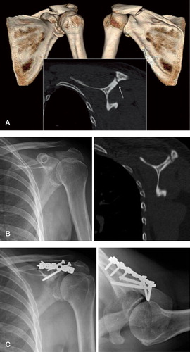
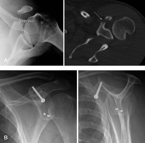
Figure 4 A. AP radiograph (left) and CT scan of the scapula (right) showing fracture of the middle coracoid with > 1 cm of displacement. B. AP (left) and lateral (right) radiographs after open reduction and internal fixation using a cortical lag screw.