Figures & data
Figure 1. Reference position. The global coordinate system is fixed to the cage (illustrated here at floor level). At the reference examination the two body-fixed coordinate systems (one scapular one humeral) are aligned to the cage system.
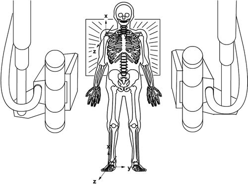
Figure 2. During motion the two body-fixed coordinate systems follow the motion of the bones which is illustrated here with changed position of the humeral coordinate system.
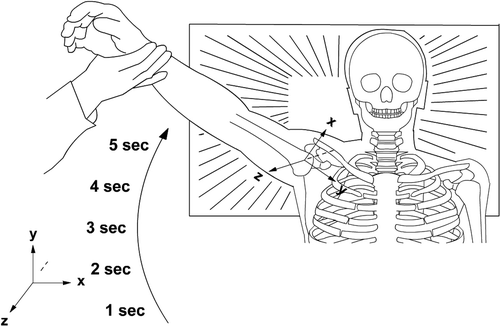
Figure 3. Simplified sketch to illustrate absolute or global motions (top) and relative humeral motions (bottom). Absolute humeral motions include changes of position caused by scapular and trunk motions whereas relative motions do not.
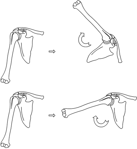
Table 1. Combined spine, trunk, scapular and humeral motions (absolute motions) at increasing degrees of motions inside the glenohumeral joint (relative motion). Recorded values and distribution between the absolute and relative glenohumeral motions in percent are presented at active abduction
Tabel 2. Combined spine, trunk, scapular and humeral motions (absolute motions) at increasing degrees of motions inside the glenohumeral joint (relative motions) Recorded values and distribution between the absolute and relative glenohumeral motions in percent are presented at passive abduction
Tabel 3. Proximal translation of the humeral head in combination with active spine, trunk, scapular and humeral motions (absolute motion) at increasing degrees of motion inside the glenohumeral joint (relative motion). Recorded values in mm proximal translations of the humeral head are presented
Tabel 4. Proximal translation of the humeral head in combination with passive spine, trunk, scapular and humeral motions (absolute motion) at increasing degrees of motion inside the glenohumeral joint (relative motion). Recorded values in mm proximal translations of the humeral head are presented. At 55 degrees of relative motion there are missing observation at absolut proximal translations in patients and the control group.
Figure 8. Angular velocity during active abduction. Patients with impingement versus the control group. Mean SE (p = 0.4).
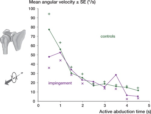
Figure 9. Angular velocity during passive abduction. Patients with impingement versus the control group. Mean SE (p = 0.7).
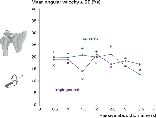
Figure 10. Translation velocity during active abduction. Patients with impingement versus the control group. Mean SE (p = 0.4).
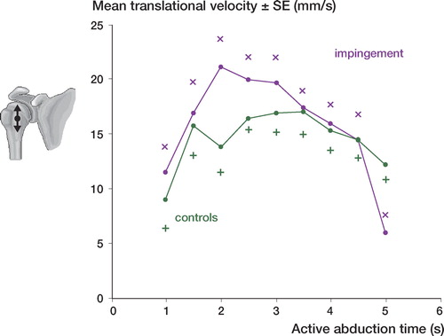
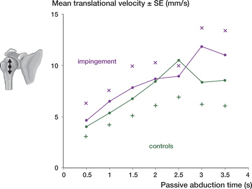
Figure 11. Translation velocity during passive abduction. Patients with impingement versus the control group. Mean SE (p = 0.5).