Figures & data
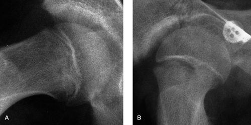
Figure 1. A. The right hip of a 13-year-old boy, showing a large epiphyseal tubercle. B. This is contrasted with the right hip of a 12-year-old boy, showing a fairly small epiphyseal tubercle, which nevertheless still has a stabilizing effect on the developing SUFE.
The various children selected from routine orthopedic clinics
Figure 2. A. An AP CT scanogram of the left hip of a 14-year-old boy showing a well-developed epiphyseal peg. B. A similar CT scanogram of the right hip of a 13-year-old boy who has a poorly defined peg that appears to be showing signs of loosening.
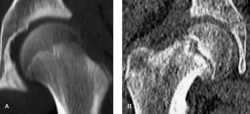
Figure 3. AP (A) and axial (B) CT scanograms of the right hip of the 13-year-old boy whose radiograph is shown in , now revealing an epiphyseal peg that seems to be a rather loose fit in the metaphyseal socket.
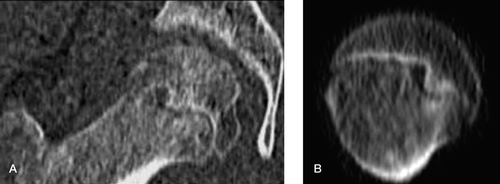
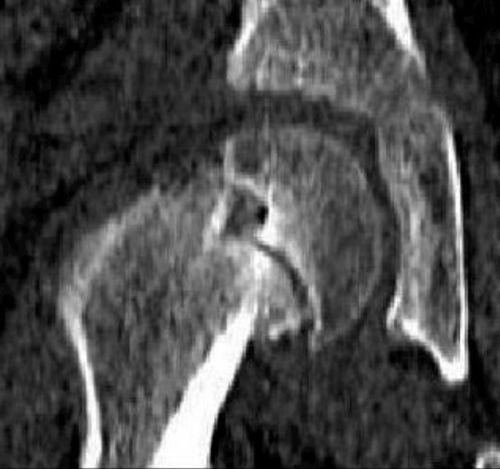
Figure 4. AP CT scanogram of the right hip of a 13-year-old boy, showing a well-developed epiphyseal tubercle that is clearly displacing out of the metaphyseal socket as an acute-on-chronic slip develops.