Figures & data
Summary of the numbers of bursae mentioned and described in all monographs and official anatomical terminologies discussed
Figure 1. Andreas Svane Dick Synnestvedt.
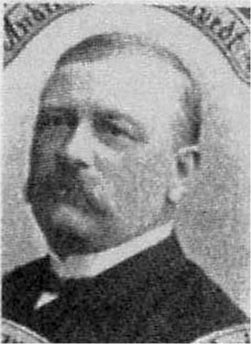
Figure 2. Title page of Synnestvedt's monograph.
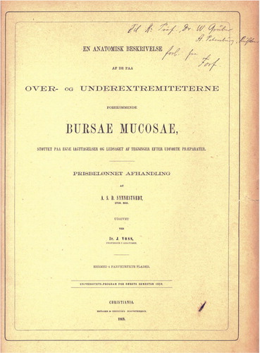
Figure 3. An example of the bursae in the shoulder region. 3: subscapular bursa; 12: subtendinous bursa of teres major.
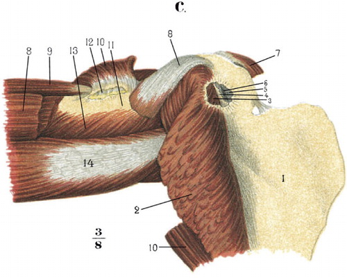
Figure 4. b. Index finger: 3: subcutaneous mucous bursa. c. 1–3: metacarpals II–IV; 8: intermetacarpophalangeal bursae.
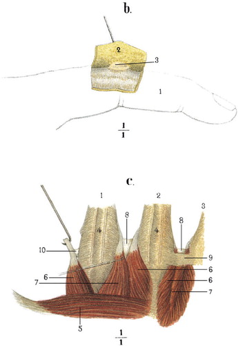
Figure 5. Depiction of same postgenual bursae. 11: medial subtendinous bursa of gastrocnemius; 13: semimembranoso-gastrocnemial bursa; 18: semimembranous bursa, prolongating in front of and behind the anterior part of semimembranosus.
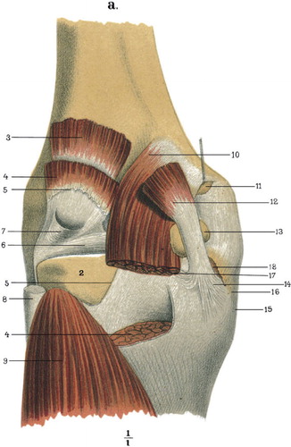
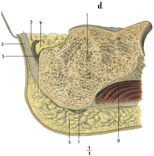
Figure 6. Sagittal section through the heel region. 2: calcaneal tendon; 3: adipose tissue, covering the superior wall of the bursa; 4: adipose fold, roaming freely in the bursa; 5: retrocalcaneal bursa; 7: subcalcaneal bursa.