Figures & data
Figure 1. Innervation of soleus muscle and vascular supply. Arrowheads: lateral border of soleus muscle (SO); arrow: neurovascular bundle coming from the gastrocnemius muscle (GM) and entering at the medial border of the SO, the tip of the arrow indicating the soleus nerve; PE: peroneal muscles. The asterisk shows the Achilles tendon.
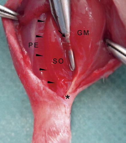
Figure 2. Time course of muscle function. Contraction forces were normalized within individuals to the forces of uninjured control muscles.
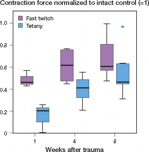
Figure 3. Time course of T1 and T2 signals after crushing of soleus muscle. Axial scans of the left lower leg of rat 2 in the MRI group, 1, 4, and 8 weeks after trauma. The asterisk shows the tibia and the arrowhead shows the fibula. Arrows: soleus muscle.
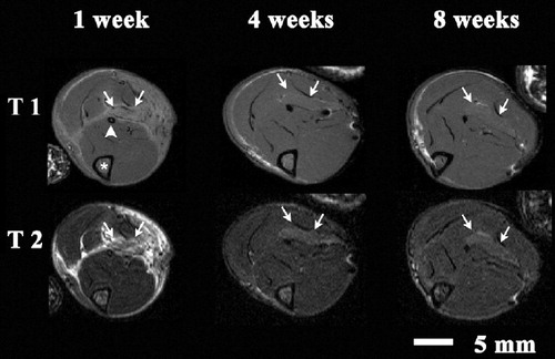
Figure 4. Time course of the differences in the grayscale values of soleus muscles and uninjured anterior tibial muscles 1, 4, and 8 weeks after soleus trauma in T1- and T2-weighted sequences. P-values: 1 week vs. 4 weeks, T1 + T2: p < 0.001; 1 week vs. 8 weeks, T1 + T2: p < 0.001; 4 weeks vs. 8 weeks, T1 and T2: p = 0.22 (ns) and p = 0.02, respectively.
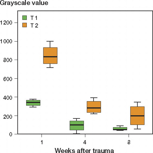
Figure 5. Longitudinal cryo-sections of crush-injured soleus muscles stained with Picrosirius red. a. Overview showing the angular uninjured region at the middle of the section (from day 1 after injury). The insertion of the neurovascular bundle is indicated by an arrow. b. Detail of panel a showing ruptured myofibers as evidence of shearing-type injury. c. Detail of panel a showing swollen, prenecrotic myofibers as evidence of in situ necrosis happening in parallel to the latter type of injury. d. Overview of the uninjured zone and the distal crush zone of a soleus muscle from day 2 after crush injury. e. Detail of panel d showing the border zone of injured and uninjured muscle with an interstitial inflammatory reaction. f. Myofibers in the crush zone surrounded by inflammatory cells in loose connective tissue.

Figure 6. Histological staining of soleus muscles 4 days and 1, 4, and 8 weeks after crush injury. a. Cryo-section with HE staining of cross-sectioned myofibers surrounded by inflammatory cells on day 4 after injury. b. Paraffin section with HE staining of regenerating myofibers showing loose deposition of connective tissue with a high degree of cellularity and regenerating muscle fibers one week after trauma. c. Paraffin section with HE staining showing dense connective tissue with local scar formation and interstitial fibrosis 4 weeks after injury. d. Cryo-section with Sirius red staining: an overview of the injured soleus muscle 8 weeks after trauma showing typical interstitially pronounced fibrosis in the distal and proximal crush zone. e. Detail of panel d showing cross-sectioned myofibers surrounded by collagenous fibrotic tissue. f. Detail of panel d showing (longitudinally) sections of myofibers and interstitial fibrosis.
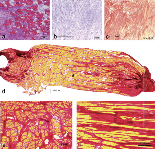
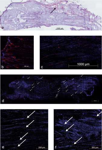
Figure 7. a. Factor VIII staining of a longitudinally sectioned soleus muscle 4 weeks after crush injury, showing the distribution of vessels in the injury zones. b. Cryo-section with an immunohistochemical stain for nestin in a healthy soleus muscle (counterstain: DAPI), myotendinous transition zone. c. Staining for nestin in a longitudinally sectioned soleus muscle (counterstain: DAPI). Nestin immunoreactivity was dispersed throughout the muscle and could be found not only at the tips of regenerating myofiber stumps but also at the lateral aspects of the fibers. d. Overview of a-bungarotoxin staining of a longitudinally sectioned soleus muscle (counterstain: DAPI). The white arrows indicate multiple, newly developed neural endplates in the proximal and distal crush zones of the muscle. e. Detail of panel d depicting a linear distribution of endplates in the uninjured region. f. Detail of panel d depicting endplates within the crushed area.
Relative amount of collagenous connective tissue over time referenced to the whole area of the muscle sections in percent (SD)