Figures & data
Table 1. Age at primary disease, type of infection, age at trochanter arthroplasty, and secondary operations
Figure 1. Patient no. 1. A. Preoperatively, showing no femoral head on the right side and the femur positioned considerably more proximally than normal. A valgization osteotomy had been performed previously. A valgization osteotomy had also been performed in the left hip. B. Postoperatively, following transposition of the greater trochanter into the acetabulum on the right side. The subtrochanteric osteotomy was fixated with a Steinman pin. C. 24 years postoperatively, showing a spherical femoral head well covered by a congruent acetabular roof. The cartilage thickness seems adequate. Note the steep femoral neck and the low medial femoral head offset. Severe osteoarthritic changes can be seen in the left hip.
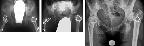
Figure 2. Patient no 2. A. Preoperatively, showing a luxated hip without any signs of a femoral neck and femoral head in the left hip. B. After trochanter arthroplasty and subtrochanteric osteotomy fixated with Steinman pins. Note severe acetabular dysplasia. C. 21 years after trochanter arthroplasty. The femoral head is flattended and poorly covered by the acetabulum. Osteoarthritic changes are present.
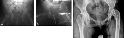
Figure 3. Patient no 3. A. Preoperatively, showing a totally luxated hip. B. 18 months after arthroplasty, the osteotomy had been fixated with a plate. The arrow points to a tiny ossification center in the new femoral head. C. 6 years after arthroplasty. Note the growth plate below the transposed greater trochanter. D. 6 years postoperatively. The new head is nearly spherical and well covered by a congruent acetabular roof.
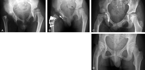
Figure 4. Patient no 4. A. Preoperatively, showing a luxated hip without any signs of a femoral head. Note the severe dysplasia of the acetabulum. B. 15 years after operation, showing that the new femoral head, which is nearly spherical, is poorly covered by a neoacetabulum above a severely dysplastic acetabulum. Osteoarthritis has developed. C. After insertion of a total hip prosthesis.
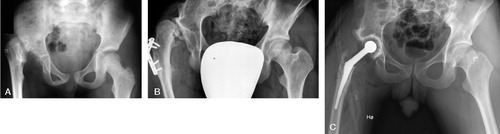
Table 2. Findings at the final follow-up examination