Figures & data
Figure 1. The TFCC is forced into rupture (specimen 4). The ulnar head is bald. Note the ECU tendon sheath torn out of its groove in the distal ulna. US: ulnar styloid with a type-1 fracture; bold arrow: longitudinal force applied to the forearm.
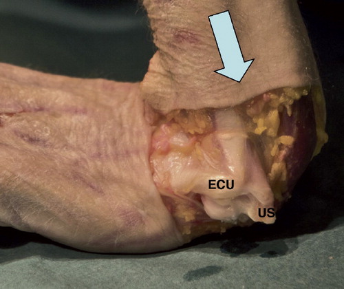
Figure 2. Classification of ulnar styloid fractures. Palmar view. Type 1: distal to the base where superficial horizontal fibers of the TFCC insert, as well as the dorsal UT ligament (Sasao et al. Citation2003). Type 2: base fracture; goes through a line perpendicular to the ulnar shaft and into the proximal limitation of the fovea, but does not involve the articular surface of the ulnar head. Type 3: proximal to a Type 2 fracture. Arrow: ECU tendon sheath.
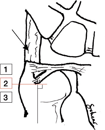
Figure 3. Sequence of displacement of the distal radius fragment in specimen 4. A. Before TFCC disruption. B. Maximum displacement. C. Pressure released.
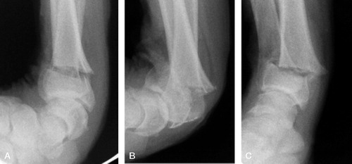
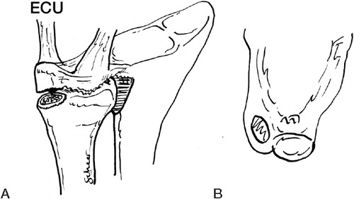
Figure 4. Type-1 foveal TFCC fiber disruption. A. Oblique palmar view. A hole is visible in the palmar capsule and foveal fibers are disrupted. Bold arrow: tension of the soft tissues in the direction of the UC, UT, and UL ligaments. B. Transverse view. Small arrow: the dorsal separation between the ECU tendon sheath and the TFCC.
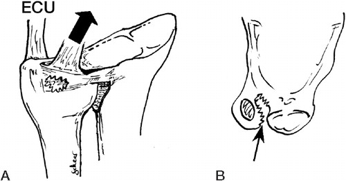
Figure 5. Type-2 foveal TFCC fibre disruption. A. Oblique palmar view. Complete disruption of the palmar capsule with either a sagittal rupture of the superficial fibers of the radioulnar ligaments (upper panel) or an ulnar styloid fracture of type 1 (lower panel). B. Transverse view. Arrow: the dorsal separation between the ECU tendon sheath and the TFCC.
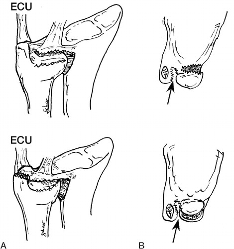
Figure 6. Fracture through the base of the ulnar styloid (type 2). A. Oblique palmar view. The TFCC was displaced together with the ulnar styloid fragment. B. Transverse view. Note the absence of dorsal separation between the ECU tendon sheath and the TFCC.