Figures & data
Figure 1. Mild adverse reaction to metal debris. Sagittal T2W MR through the femoral stem (S) of a Corail total hip replacement demonstrating mild periprosthetic disease. A small fluid-filled cavity (asterisk) surrounding the neck of the prosthesis is encapsulated by a thick, ragged low-signal rim (white arrow).
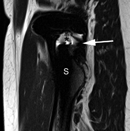
Figure 2. Moderate adverse reaction to metal debris. A sagittal T2W MR positioned just medial to the acetabular cup demonstrates moderate periprosthetic disease with a large cystic collection, demarcated by a low signal wall (black arrow), and filled with debris (white arrow) extending proximally in the line of the iliopsoas bursa. The relatively thick low signal wall and the debris are not typical of conventional iliopsoas bursae.
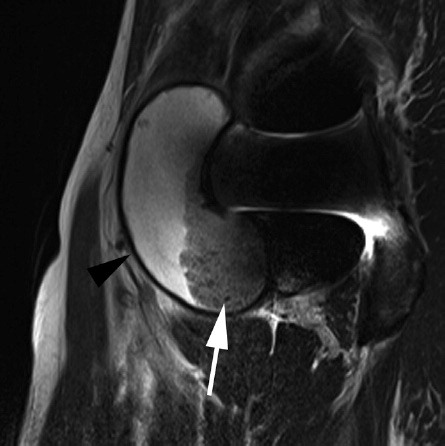
Figure 3. Severe adverse reaction to metal debris. Coronal T1W MR through the mid-coronal plane of the femoral head (black arrows indicate the medial wall of the acetabulum), demonstrating severe periprosthetic disease with bone marrow replacement in the acetabular roof (white arrow).
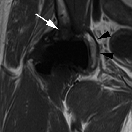
Table 1. Summary of metal artifact-reduction (MAR) MRI findings
Table 2. Metal artifact-reduction MRI findings in relation to potential risk factors for metal debris-related reactions
Table 3. Summary of revisions
Table 4. Patient-related outcome in relation to MAR MRI findings