Figures & data
Figure 1. Case 1. The typical coxa valga due to coalescence (arrow) on a standard AP view (A) and T1-weighted coronal MRI of the pelvis during growth at the age of 13 years (B). On the Lauenstein view, the head-neck offset is small (C).
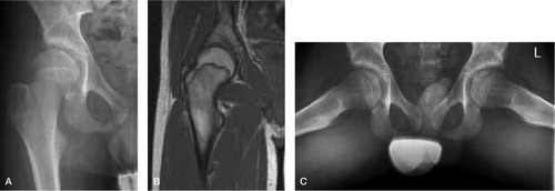
Figure 2. Case 2. A and B. Marked coalescence and almost absent anterior head-neck offset on standard AP (A) and Lauenstein (B) views at 14 years of age. C. AP pelvic view at 16 years of age.
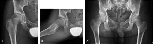
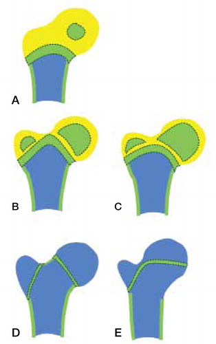
Figure 3. Schematic representation of the growing human hip region, showing the primary femoral and secondary capital and greater trochanteric ossification centers and proximal diaphyseal growth plate at ages of 1 year (A), 2–4 years (B, C), and > 4 years (D, E). Panels A, B, and D represent normal development, showing isthmic interruption of the chondroepiphysis prior to trochanteric osseous expansion, normal orientation of the capital growth plate, and normal head-neck offset. Panels C and E represent persistence of the isthmic part of the chondroepiphysis with subsequent coalescence of expanding secondary ossification centers, horizontal orientation of the capital growth plate, and reduced head-neck offset.