3,439
Views
48
CrossRef citations to date
0
Altmetric
Research Article

Isolated fractures of the greater tuberosity of the proximal humerus

A long-term retrospective study of 30 patients

, , , , , , , & show all
Pages 714-720 | Published online: 06 Sep 2011

Figures & data

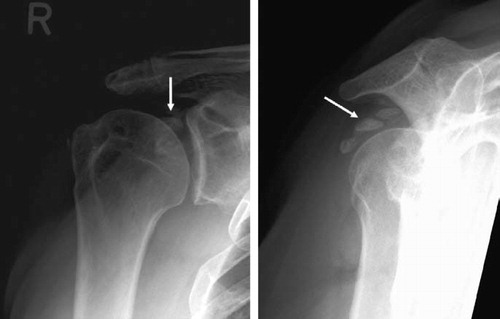
Figure 1. The left shoulder of a 64-year-old patient (no. 3, ), who suffered an anterior glenohumeral dislocation of the left shoulder with an avulsed greater tuberosity.

Figure 1. The left shoulder of a 64-year-old patient (no. 3, Table 3), who suffered an anterior glenohumeral dislocation of the left shoulder with an avulsed greater tuberosity.

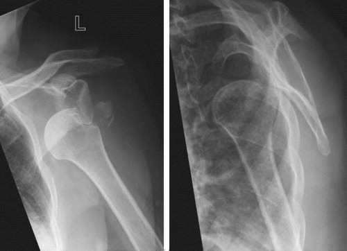
Figure 2. Patient no. 9 () before and after open reduction and internal fixation with cannulated screws of a moderately displaced fracture of the greater tuberosity (white arrows). An MRI scan illustrates the fracture line (white arrows).

Figure 2. Patient no. 9 (Table 3) before and after open reduction and internal fixation with cannulated screws of a moderately displaced fracture of the greater tuberosity (white arrows). An MRI scan illustrates the fracture line (white arrows).

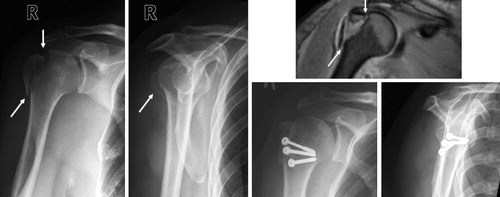
Figure 3. Patient no. 6 () before and after open reduction and internal fixation of greater tuberosity fragments with major displacement (white arrows). The multiple fragments were fixed with a screw and a tension wire.

Figure 3. Patient no. 6 (Table 3) before and after open reduction and internal fixation of greater tuberosity fragments with major displacement (white arrows). The multiple fragments were fixed with a screw and a tension wire.

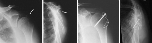
Figure 4. Patient no. 18 () before and after open reduction and plate fixation of a moderately displaced fracture of the greater tuberosity (white arrows).

Figure 4. Patient no. 18 (Table 3) before and after open reduction and plate fixation of a moderately displaced fracture of the greater tuberosity (white arrows).

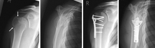
Table 1. Overview of clinical evaluation considering fragment displacement

Table 2. Active ROM at the time of follow-up examination

Table 3. Synopsis of clinical and radiographic evaluation of patients with isolated greater tuberosity fractures of the humerus

Figure 5. Radiographs of the right shoulder illustrating severe periarticular calcification (white arrows) of a patient (no. 30, ) who was treated operatively with ORIF with screws for a major displaced fracture of the greater tuberosity. After radiographic healing, the metal was removed.

Figure 5. Radiographs of the right shoulder illustrating severe periarticular calcification (white arrows) of a patient (no. 30, Table 3) who was treated operatively with ORIF with screws for a major displaced fracture of the greater tuberosity. After radiographic healing, the metal was removed.