Figures & data
Figure 1. Synovial tissue necrobiosis. (A) Villous proliferations without stainable synoviocytes, blood vessel walls, stromal cells, and inflammatory infiltrates (at top right) were characteristic of villous necrobiosis. (B) Band-like acellular areas of the synovial surface (upper half) showing scattered ghost cells were a typical finding in synovial surface necrobiosis. Their maximal extent vertically was measured digitally and recorded in each case. (C) Similar acellular areas (at lower right) in deeper periarticular soft tissue without any contact with the synovial surface in this specimen were referred to as deep necrobiosis. (Hematoxylin and eosin).
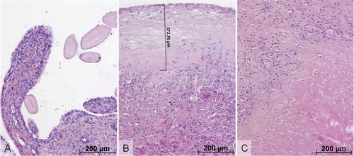
Figure 2. Immunohistochemistry with antibody to CD3 antigen was performed in order to identify T-lymphocytes within the synovial soft tissue. In this particular case, both perivascular (mid right) and diffuse CD3+ lymphocytes were detected. Polyethylene wear particles were apparent under polarized light microscopy in several foreign body giant cells and histiocytes. (Embedding in paraffin wax; microscopic analysis under polarized light; immunohistochemical reaction with CD3 antibody).
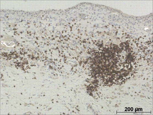
Table 1. Patterns of necrobiosis in the study cohort (Fisher's exact test)
Table 2. Density of T-lymphocyte subpopulations in the study cohort (Mann-Whitney U test). Values are median (IQR)