Figures & data
Figure 1. Region of interest used for µCT analysis. Frontal and transverse sections of the proximal tibia (epiphysis removed) from saline-treated (A) and Scl-Ab-treated (B) weight-bearing animals. A 1.5-mm long region in the metaphysis was chosen (dashed lines). In this region, only the trabecular bone was evaluated (white trace). This region was situated away from the screw (*) so that it would not be influenced by any bone formation around the screw.
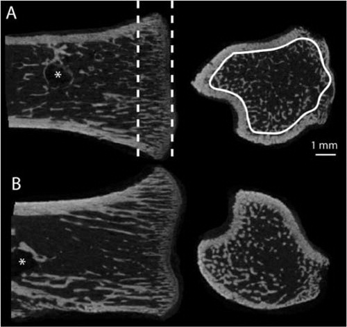
Figure 2. Effect of treatment with anti-sclerostin antibody (Scl-Ab) on screw fixation. A. Peak pull-out force. B. Pull-out energy. C. Stiffness. Compared to controls, Scl-Ab treatment significantly increased peak force (a: p = 0.01; b: p < 0.001) and energy (c: p = 0.02; d: p < 0.001) both with and without loading. Stiffness was significantly increased in an unloaded setting (e: p = 0.03). Unloading had a large effect on screw fixation, causing a large reduction in peak force (p < 0.001).
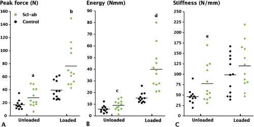
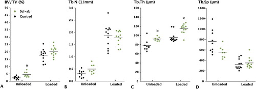
Figure 3. Effect of anti-sclerostin antibody (Scl-Ab) on trabecular bone volume in the proximal tibia, as measured by µCT. A. Bone volume fraction. B. Trabecular number. C. Trabecular Thickness. D. Trabecular separation. Botox treatment significantly reduced BV/TV, Tb.N, and Tb.Th compared to loaded controls (p < 0.001). Antibody treatment increased BV/TV both in the loaded and unloaded tibia, but only the increase in the unloaded tibia was significant (a: p = 0.03). Trabecular thickness was also significantly increased, however (b, c: p < 0.001).