Figures & data
Primer sequences and melt temperatures (Tm) used in the experiment
Figure 1. Gene expression in unfractured rib and in 7, 14, and 21 day rib fracture callus of rat (n = 7 biological replicates), normalized to GAPDH relative to normal intact rib. Expression of αSMA mRNA (A), h1CaP mRNA (B), EDA-fibronectin mRNA (C), and connexin-43 mRNA (D) all increased at 7 days post-fracture compared to unfractured rib (*p < 0.05, **p < 0.01). By 21 days post-fracture, expression of both αSMA mRNA and connexin-43 mRNA in callus was less than 7 day expression (***p < 0.001, **p < 0.01, respectively) and was similar to the levels in unfractured rib. The expression of non-muscle myosin mRNA (E) and h-caldesmon mRNA (F) was not significantly different in unfractured rib and callus at any time point.
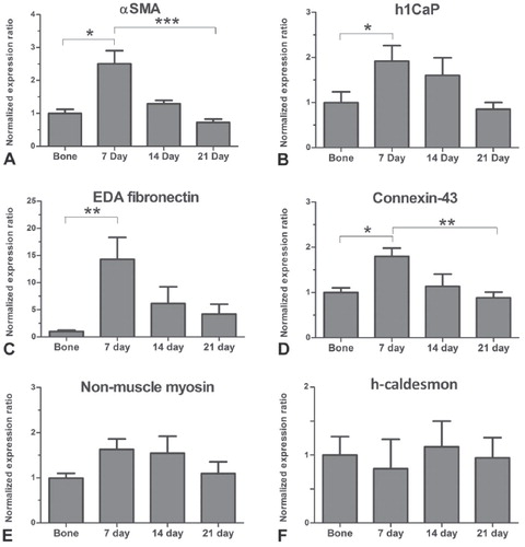
Figure 2. A low-power micrograph of a toluidine blue stained histological section of a rat rib fracture site after 7 days, showing a significant amount of soft-tissue collagenous matrix containing osteoprogenitor cells (arrows) linking together two ends (E) of fractured rib. Immunohistochemical analyses were performed in these fibrous tissue regions. Newly formed cartilage (C) and trabecular bone (B) within the callus are also evident. (M: skeletal muscle). Magnification: ×25.
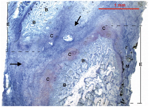
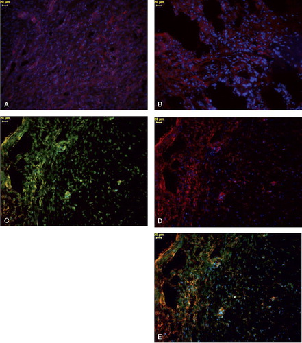
Figure 3. Immunohistochemistry of fibrous callus linking fracture ends in a rat rib fracture after 7 days. Panels C–E show immunostaining of the same callus section. Cell nuclei for panels A–E were stained with DAPI (blue).
A.Osteoprogenitor cells show consistent but moderate CaP-LI (indicated by red fluorescence).
B.There is intense EDA-fibronectin-like immunoreactivity (LI) (indicated by red fluorescence) in some regions of callus; other areas show only moderate EDA-fibronectin-LI immunostaining.
C.OB-cadherin-LI in osteoprogenitor cells (indicated by yellow-green fluorescence). OB-cadherin-LI is particularly intense in high-density fibrous regions (left of picture). Not all cells are OB-cadherin-positive, however.
D.Osteoprogenitor cells to the left contain significant αSMA-LI (indicated by red fluorescence), but the cells to the right show only moderate αSMA-LI.
E.A sequentially immunostained section for αSMA (red fluorescence) and OB-cadherin (yellow-green fluorescence). Co-localization of these two proteins by immunohistochemistry resulted in a combined brown-orange fluorescence, which is mainly located in osteoprogenitor cells to the left of this micrograph. Note that some cells only show either αSMA-LI or OB-cadherin-LI. Magnification: ×200; scale bars = 20 μm.