Figures & data
Figure 1. Peak force (N) at tensile testing of transected tendons that were allowed to heal either with voluntary loading or with muscle paralysis induced by Botox.
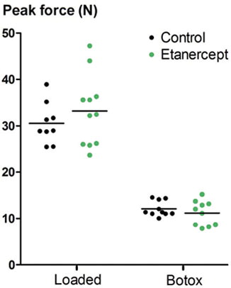
Table 1. 95% confidence limits for the difference between means for mechanical properties of transected tendons (etanercept minus control). Values are expressed as percent of control (percent change from etanercept). The tendons were allowed to heal with voluntary loading or with muscle paralysis induced by Botox
Figure 2. Data from pull-out testing of screws 14 days after insertion in the proximal tibial metaphysis. Etanercept was given early, late, or continuously over the 14-day period.
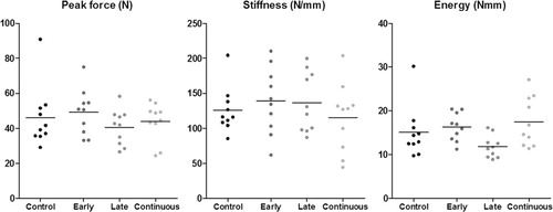
Table 2. 95% confidence limits for the difference between means for screw fixation (the etanercept groups combined minus controls). Values are expressed as percent of control (percent change from etanercept)
Figure 3. Ossicles induced by BMP-2 in a collagen scaffold. Row A: control. Row B: continuously etanercept-treated. Column I: raw image. Column II: low threshold (ossicle size). Column III: high threshold (estimate of bone density).
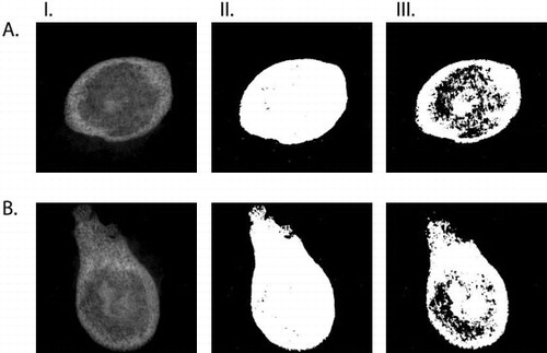
Figure 4. Radiographic data from rh-BMP 2 implants, with two different gray-scale thresholds for blind examination. Ossicle size (A) was measured with a low threshold, and “bone area” (B) with a higher threshold, giving a visual impression of being related to bone density. For comparison, both estimates are presented in mm2. Ossicle size was larger with etanercept treatment (pooled; p = 0.02).
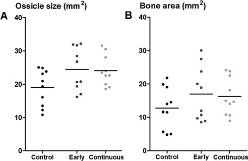
Table 3. 95% confidence limits for the difference between means for radiographic data describing ossicles induced by BMP-2 (both the etanercept groups combined minus controls). Values are expressed as percent of control (percent change from etanercept)
Table 4. Histological scoring of endochondral ossification: median (min–max). The scale goes from 0 (no prevalence) to 3 (high prevalence). The p-values are from Kruskall-Wallis test