Figures & data
Radiographic measurement of the lateral tibiocalcaneal angle in each group of patients. A. A lateral tibiocalcaneal angle of 82° before PAT in a 3-month-old infant. B. The same infant at 15 months of age; the lateral tibiocalcaneal angle was 67°. This child was assigned to group 1. C. A lateral tibiocalcaneal angle of 118° before percutaneous Achilles tenotomy in a 3-month-old infant. D. The same infant at 12 months of age; the lateral tibiocalcaneal angle was 96° before selective soft tissue release was performed to treat the residual equinovarus deformity. This child was assigned to group 2.
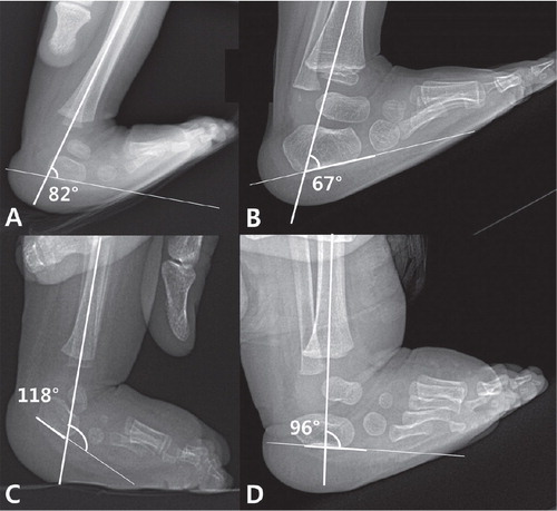
Table 1. Characteristics of the 10 clubfeet in 7 patients with residual clubfoot deformities
Table 2. Demographics of groups 1 and 2. Values are mean (SD)
Table 3. Clinical scores of groups 1 and 2. Values are least-squares mean (SE) [95% CI]
Table 4. Radiographic data for groups 1 and 2 at the time of PAT. Values are least-squares means (SE) [95% CI]
Table 5. Degree of correlation between Pirani score at the last follow-up examination and the radiographic parameters measured at the time of PAT. All values are correlation coefficient (p-value)
Table 6. Radiographic results of the 50 clubfeet that were evaluated by 2 observers at the time of PAT