Figures & data
Figure 1. Gap-implant in the proximal tibia. Radiograph taken post mortem.
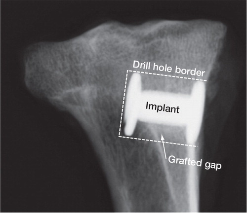
Figure 2. Experimental porous-coated titanium 2.5 mm gap implant with measurements.
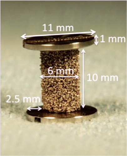
Table 1. Allograft wash
Figure 3. Bone graft of the 2 treatment groups.
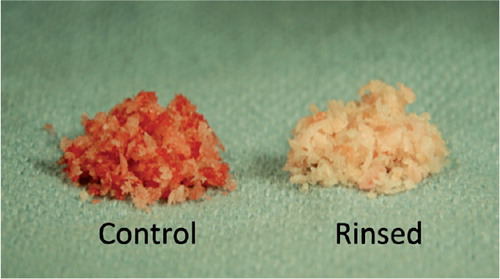
Figure 4. Histology: representative histological sections from the two groups. The sections shown are from the two implants inserted in the same animal. Porous-coated Ti implants are surrounded by a 2.5-mm concentric defect. The sections are cut parallel to the long axis of the implant. Left-hand panels (control): fibrous (FIB) tissue at the implant (IMPL) surface. Right-hand panels (rinsing): new bone (NB) on graft (AG) remnants.
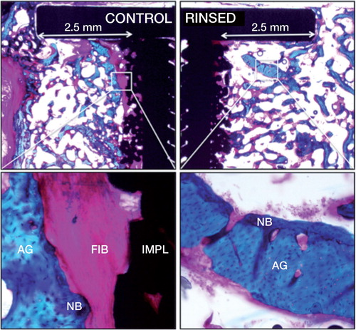
Table 2. Biomechanical results
Table 3. Histomorphometrical analysis: tissue area fraction on implant surface and tissue volume fraction in the gap (ingrowth)