Figures & data
Figure 1. Immunohistochemistry of supraspinatus muscle, showing MHC1+ (left panels) and MHC2+ fibers (right panels). A and B. Serial sections were stained with the 2 relevant antibodies (see Methods for details). C and D. The same pixel selection algorithm was used to select positive staining (orange/red) and negative staining (blue). E and F. Higher-magnification view of area shown in A and B. Note the generally non-overlapping pattern of MHC1 and MHC2 immunostaining. The vast majority of myofibers have been stained with one of the antibodies, but not both, while a very small minority have not been stained with either.
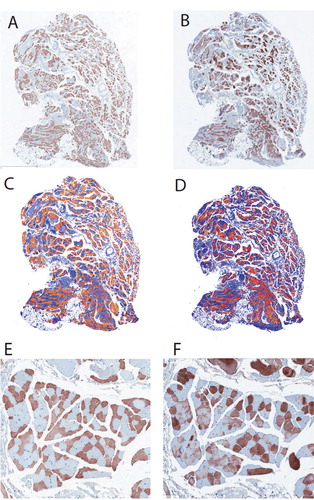
Figure 2. Sample fiber size distribution of type 1 and type 2 myofibers in the supraspinatus muscle of patients with partial-thickness rotator cuff tears (PTRCT, top panels) or full-thickness rotator cuff tears (FTRCT, bottom panels). Note the smaller size (a leftward shift in myofiber histogram) of both myofiber types in the FTRCT patients.
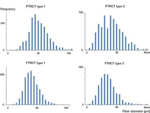
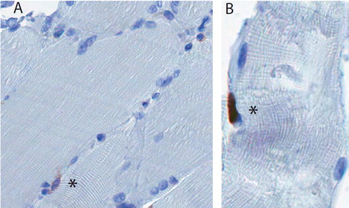
Figure 3. A. Satellite cells were defined as CD56+ cells at the periphery of the myofiber (marked with an asterisk). B. A Ki67+ cell (asterisk), which appears to lie outside the sarcolemma, in the expected location of a satellite cell.